Fraud Blocker Skip to main content

Home > All Images > 1998 > August > 6 Aug 1998

Images Dated 6th August 1998

Choose a picture from our collection for your Wall Art and Photo Gifts

33 Items

Background imageImages Dated 6th August 1998: TV Programme Take the High Road Deborah McCallum August 1998

TV Programme Take the High Road Deborah McCallum August 1998

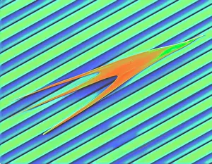
Background imageImages Dated 6th August 1998: Coloured SEM of laptop screen foil surface

Coloured SEM of laptop screen foil surface
Laptop screen foil. Coloured scanning electron micrograph (SEM) of the surface of a brightness- enhancing foil used to make laptop personal computer screens look brighter

Background imageImages Dated 6th August 1998: Mr Dennis Russell of Walsall, with one of his Gurning faces. 8th June 1998

Mr Dennis Russell of Walsall, with one of his Gurning faces. 8th June 1998

Background imageImages Dated 6th August 1998: Tom Read former SAS Soldier nursed back to sanity by his wife Livvy Read after the Gulf

Tom Read former SAS Soldier nursed back to sanity by his wife Livvy Read after the Gulf war had driven him mad August 1998

Background imageImages Dated 6th August 1998: Fly on the wall feature on the Swallow Hotel, Stockton. General Manager Paul Morrison

Fly on the wall feature on the Swallow Hotel, Stockton. General Manager Paul Morrison. 6th August 1998

Background imageImages Dated 6th August 1998: Vauxhall Corsa red August 1998 Man leaning on bonnet

Vauxhall Corsa red August 1998 Man leaning on bonnet

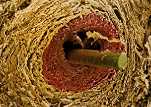
Background imageImages Dated 6th August 1998: Coloured SEM of a section through a hair follicle

Coloured SEM of a section through a hair follicle
Hair follicle. Coloured scanning electron micrograph (SEM) of a cross-section through a hair follicle below the skin surface

Background imageImages Dated 6th August 1998: Fly on the wall feature on the Swallow Hotel, Stockton. General Manager Paul Morrison

Fly on the wall feature on the Swallow Hotel, Stockton. General Manager Paul Morrison talking to the Head Chef. 6th August 1998

Background imageImages Dated 6th August 1998: Scene from STV programme Take The High Road August 1998

Scene from STV programme Take The High Road August 1998

Background imageImages Dated 6th August 1998: England v South Africa 5th Test Headingley Aug 1998 Mark Butcher batting during his

England v South Africa 5th Test Headingley Aug 1998 Mark Butcher batting during his century innings

Background imageImages Dated 6th August 1998: England v South Africa cricket 5th Test Headingley Aug 1998 Mark Butcher leaving

England v South Africa cricket 5th Test Headingley Aug 1998 Mark Butcher leaving the crease after being bowled by Shaun Pollock

Background imageImages Dated 6th August 1998: Mr Dennis Russell of Walsall with another of his gurning th June 1998

Mr Dennis Russell of Walsall with another of his gurning th June 1998

Background imageImages Dated 6th August 1998: Ken Livingstone August 1998 at London Zoo to promote the new Web Of Life building being

Ken Livingstone August 1998 at London Zoo to promote the new Web Of Life building being constructed - holding Cuddles - a Boa Constrictor Snake

Background imageImages Dated 6th August 1998: England v South Africa 5TH Test Headingley Aug 1998 Mark Butcher celebrating

England v South Africa 5TH Test Headingley Aug 1998 Mark Butcher celebrating after scoring a century

Background imageImages Dated 6th August 1998: Ken Livingstone August 1998 at London Zoo to promote the new Web Of Life building being

Ken Livingstone August 1998 at London Zoo to promote the new Web Of Life building being constructed - holding Cuddles - a Boa Constrictor Snake

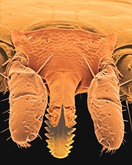
Background imageImages Dated 6th August 1998: Coloured SEM of the head of a tick, Ixodes sp

Coloured SEM of the head of a tick, Ixodes sp
Lyme disease tick. Coloured scanning electron micrograph (SEM) of the head of a tick (Ixodes sp.), the principal vector of Lyme disease

Background imageImages Dated 6th August 1998: Coloured SEM of a tick (Ixodes sp. ) in human skin

Coloured SEM of a tick (Ixodes sp. ) in human skin
Feeding tick. Coloured scanning electron micrograph (SEM) of a tick (Ixodes sp. ) feeding head- down in human skin

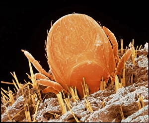
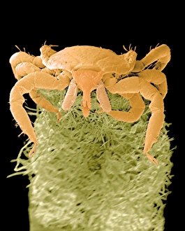
Background imageImages Dated 6th August 1998: Coloured SEM of a tick, Ixodes sp. on plant leaf

Coloured SEM of a tick, Ixodes sp. on plant leaf
Lyme disease tick. Coloured scanning electron micrograph (SEM) of a tick (Ixodes sp.) on a plant leaf

Background imageImages Dated 6th August 1998: Coloured SEM of a tick (Ixodes sp. ) in human skin

Coloured SEM of a tick (Ixodes sp. ) in human skin
Feeding tick. Coloured scanning electron micrograph (SEM) of a tick (Ixodes sp.) feeding head-down in human skin

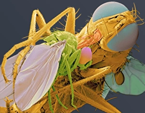
Background imageImages Dated 6th August 1998: Coenosia fly eating a drosophila

Coenosia fly eating a drosophila
Fly eating another fly. Coloured scanning electron micrograph (SEM) of a large fly (Coenosia humilis, orange) eating a smaller fruit fly (Drosophila sp.)

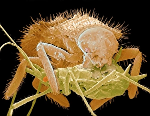
Background imageImages Dated 6th August 1998: Coloured SEM of ladybird larva eating an aphid

Coloured SEM of ladybird larva eating an aphid
Ladybird larva eating aphid. Coloured scanning electron micrograph (SEM) of a ladybird larva (Coccinella sp. orange) feeding on an aphid (pale green). The larva is holding the aphid in its jaws

Background imageImages Dated 6th August 1998: Coloured SEM of a Coenosia fly eating a Drosophila

Coloured SEM of a Coenosia fly eating a Drosophila
Fly eating another fly. Coloured scanning electron micrograph (SEM) of a large fly (Coenosia humilis, orange) eating a smaller fruit fly (Drosophila sp.)

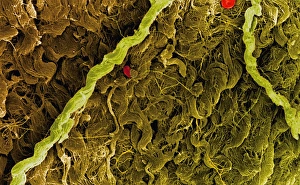
Background imageImages Dated 6th August 1998: Coloured SEM of collagen connective tissue fibres

Coloured SEM of collagen connective tissue fibres
Collagen connective tissue. Coloured scanning electron micrograph (SEM) of connective tissue formed by bundles of wavy collagen fibres. Red blood cells (red) are also seen

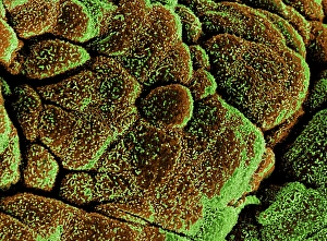
Background imageImages Dated 6th August 1998: Coloured SEM of healthy bladder epithelium

Coloured SEM of healthy bladder epithelium
Bladder epithelium. Coloured scanning electron micrograph (SEM) of healthy urinary bladder epithelium or mucosa

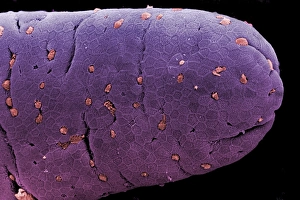
Background imageImages Dated 6th August 1998: Coloured SEM of a villus of the small intestine

Coloured SEM of a villus of the small intestine
Villus of small intestine. Coloured scanning electron micrograph (SEM) showing a healthy villus of the small intestine

Background imageImages Dated 6th August 1998: Coloured SEM of a villus of the small intestine

Coloured SEM of a villus of the small intestine
Villus of small intestine. Coloured scanning electron micrograph (SEM) showing a healthy villus of the small intestine

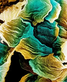
Background imageImages Dated 6th August 1998: Coloured SEM of epithelial cells in the oesophagus

Coloured SEM of epithelial cells in the oesophagus
Oesophagus epithelium. Coloured scanning electron micrograph (SEM) of the epithelium (lining) of the oesophagus. The irregularly-shaped epithelial cells (blue & yellow) have a highly folded surface

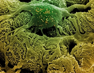
Background imageImages Dated 6th August 1998: Coloured SEM of podocytes in the human kidney

Coloured SEM of podocytes in the human kidney
Kidney glomerulus. Coloured scanning electron micrograph (SEM) of podocyte cells on a glomerulus in the kidney

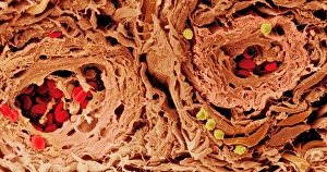
Background imageImages Dated 6th August 1998: Coloured SEM of blood vessels in the skin

Coloured SEM of blood vessels in the skin
Skin blood vessels. Coloured scanning electron micrograph (SEM) of two small blood vessels (far left and upper right) in the dermis of the skin

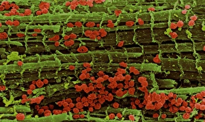
Background imageImages Dated 6th August 1998: Coloured SEM of healthy heart muscle fibres

Coloured SEM of healthy heart muscle fibres
Heart muscle. Coloured scanning electron micrograph (SEM) of healthy heart (cardiac) muscle fibres (green). Mitochondria (red) supply the muscle cells with energy

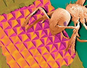
Background imageImages Dated 6th August 1998: Coloured SEM of an ant on some reflective foil

Coloured SEM of an ant on some reflective foil
Reflective material. Coloured scanning electron micrograph (SEM) of an ant on some reflective material

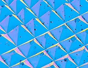
Background imageImages Dated 6th August 1998: Coloured SEM of the surface of advanced sandpaper

Coloured SEM of the surface of advanced sandpaper
Sandpaper. Coloured scanning electron micrograph (SEM) of the surface of advanced sandpaper showing the regular pyramidal arrangement on its surface

Background imageImages Dated 6th August 1998: Coloured SEM of uterus wall (early proliferation)

Coloured SEM of uterus wall (early proliferation)
Uterus (early proliferation). Coloured scanning electron micrograph (SEM) of the endometrium (lining) of the human uterus during the early proliferative stage of the menstrual cycle


All products are expertly crafted, using premium materials, tailored to your specifications and promptly shipped


© Copyright Media Storehouse, All Rights Reserved 2003 - 2025
Facebook     Pinterest     100% Payment Secure     Reviews IO