Fraud Blocker Skip to main content

Home > All Images > 2010 > November > 23 Nov 2010

Images Dated 23rd November 2010 (page 5)

589 items

Background imageImages Dated 23rd November 2010: Lime tree stem, light micrograph

Lime tree stem, light micrograph
Lime tree stem. Light micrograph of a cross-section through a three-year-old stem growing on a lime tree (Tilia europaea)

Background imageImages Dated 23rd November 2010: Muscle motor neurones, light micrograph

Muscle motor neurones, light micrograph
Muscle motor neurones. Light micrograph of dendrites (axons, black) from motor nerve cells (motor neurones) in muscle tissue (pink-purple strands)

Background imageImages Dated 23rd November 2010: Small intestine section, light micrograph

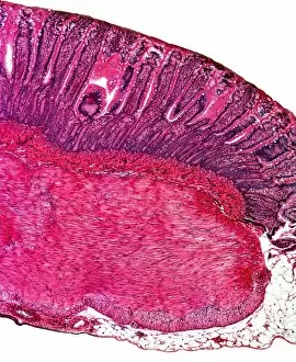
Small intestine section, light micrograph
Small intestine section. Light micrograph of a vertical section through the illeum, part of the small intestine. The upper layer (the digestive surface)

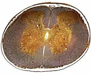
Background imageImages Dated 23rd November 2010: Spinal cord, light micrograph

Spinal cord, light micrograph
Spinal cord. Light micrograph of a cross-section through a mammalian spinal cord. The central canal (white) is surrounded by dorsal (light brown) and ventral horns (dark brown)

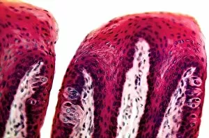
Background imageImages Dated 23rd November 2010: Tongue section, light micrograph

Tongue section, light micrograph
Tongue section. Light micrograph of a vertical section through a mammalian tongue. The surface of the tongue (across top) consists of stratified squamous epithelium (thick red-orange layer)

Background imageImages Dated 23rd November 2010: Ovarian follicle, light micrograph

Ovarian follicle, light micrograph
Ovarian follicle. Light micrograph of a transverse section through a mammalian ovary, the reproductive structure that produces the female sex cells (ova)

Background imageImages Dated 23rd November 2010: Tree growth rings, light micrograph

Tree growth rings, light micrograph
Tree growth rings. Polarised light micrograph of a cross-section through a woody stem, showing a portion of an annual ring of xylem tissue with periods of seasonal growth

Background imageImages Dated 23rd November 2010: Flatworm, light micrograph

Flatworm, light micrograph
Flatworm. Light micrograph of a Dugesia planarian flatworm. This flatworm was placed in water containing Indian ink, and the black particles have been ingested by the pharynx

Background imageImages Dated 23rd November 2010: France-Italy border at night, ISS image

France-Italy border at night, ISS image. Image taken by astronauts aboard the International Space Station (ISS) of the border between France and Italy. North is centre bottom

Background imageImages Dated 23rd November 2010: Kidney, light micrograph

Kidney, light micrograph
Kidney. Light micrograph of a vertical section through a whole kidney. The kidney is surrounded by a capsule (dark purple) under which is the cortex (deep red)

Background imageImages Dated 23rd November 2010: Pine tree stem, light micrograph

Pine tree stem, light micrograph
Pine tree stem. Polarised light micrograph of section through a young (one-year) stem from a Pinus pine tree. At top right are parenchyma cells of the cortex

Background imageImages Dated 23rd November 2010: Mollusc radula, light micrograph

Mollusc radula, light micrograph
Mollusc radula. Polarised light micrograph of the teeth on the radula ( tongue ) of the tropical mollusc Lathina rotundata. The radula is a toothed chitinous organ in the mouth of gastropods

Background imageImages Dated 23rd November 2010: Butterfly head, light micrograph

Butterfly head, light micrograph
Butterfly head. Light micrograph of the head of a butterfly (bottom) with its long segmented sensory antennae (centre and top)

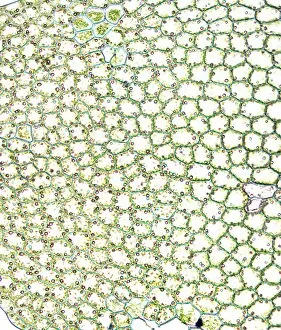
Background imageImages Dated 23rd November 2010: Liverwort leaf tissue, light micrograph

Liverwort leaf tissue, light micrograph
Liverwort leaf tissue. Light micrograph of the bifurcated tip of a leaf from a leafy liverwort (Lophocolea cuspidata). These leaves are extremely thin

Background imageImages Dated 23rd November 2010: Mammalian ovary, light micrograph

Mammalian ovary, light micrograph
Mammalian ovary. Light micrograph of a transverse section through a mammalian ovary, the reproductive structure that produces the female sex cells (ova)

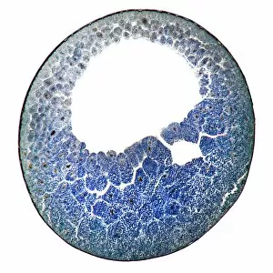
Background imageImages Dated 23rd November 2010: Developing frog egg, light micrograph

Developing frog egg, light micrograph
Developing frog egg. Light micrograph of a vertical section through a developing egg laid by a common frog (Rana temporaria temporaria). The egg has reached the blastula stage

Background imageImages Dated 23rd November 2010: Developing frog egg, light micrograph

Developing frog egg, light micrograph
Developing frog egg. Light micrograph of a vertical section through a developing egg laid by a common frog (Rana temporaria temporaria). The egg has reached the yolk plug, gastrula stage

Background imageImages Dated 23rd November 2010: Virginia creeper stem, light micrograph

Virginia creeper stem, light micrograph
Virginia creeper stem. Polarised light micrograph of a cross-section through a stem from a Virginia creeper (Parthenocissus tricuspidatus)

Background imageImages Dated 23rd November 2010: Pine tree needle, light micrograph

Pine tree needle, light micrograph
Pine tree needle. Polarised light micrograph of a cross-section through a needle from a Pinus pine tree. This leaf is needle-like in order to reduce water loss (transpiration)

Background imageImages Dated 23rd November 2010: Celery stalk, light micrograph

Celery stalk, light micrograph
Celery stalk. Light micrograph of a cross-section through a stalk from a celery plant (Apium graveolens). Red eosin dye has been used to stain the xylem tissues

Background imageImages Dated 23rd November 2010: Tree-of-heaven stem, light micrograph

Tree-of-heaven stem, light micrograph
Tree-of-heaven stem. Polarised light micrograph of a cross-section through the stem of the tree-of-heaven (Ailanthus glandulosa). Below the outer layers (red) is a ring of vascular bundles

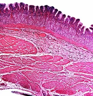
Background imageImages Dated 23rd November 2010: Stomach section, light micrograph

Stomach section, light micrograph
Stomach section. Light micrograph of a vertical section through a stomach. The upper layer (pink-purple) is the glandular mucosa

Background imageImages Dated 23rd November 2010: Skin section, light micrograph

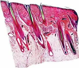
Skin section, light micrograph
Skin section. Semi-polarised light micrograph of a vertical section through human skin. The outer epidermis is the thin layer across top, with the dermis layer beneath it

Background imageImages Dated 23rd November 2010: Butterfly proboscis, light micrograph

Butterfly proboscis, light micrograph
Butterfly proboscis. Light micrograph of the coiled proboscis ( tongue ) of a butterfly. The proboscis of a butterfly consists of two half-tubes which join together in the mid-line to form a single

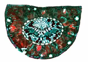
Background imageImages Dated 23rd November 2010: Aralia leaf stalk, light micrograph

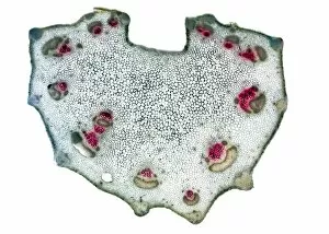
Aralia leaf stalk, light micrograph
Aralia leaf stalk. Light micrograph of a cross-section through a leaf stalk (petiole) from an Araliatree. The outer layer directly below the epidermis is a supporting collenchyma ring (red)

Background imageImages Dated 23rd November 2010: Motor neurones, light micrograph

Motor neurones, light micrograph
Motor neurones. Light micrograph of motor nerve cells (motor neurones) and their support cells. The cell bodies of the neurones (large deep purple areas) have a central nucleus

Background imageImages Dated 23rd November 2010: Sea urchin spine, light micrograph

Sea urchin spine, light micrograph
Sea urchin spine. Polarised light micrograph of a cross-section through a spine from an Echinus sea urchin. Sea urchin spines consist of supporting calcareous radial plates

Background imageImages Dated 23rd November 2010: Bracken fern rhizome, light micrograph

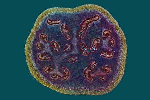
Bracken fern rhizome, light micrograph
Bracken fern rhizome. Polarised light micrograph of a cross-section through a rhizome from a bracken fern (Pteris aquilinum). The outer layer (yellow-green) consists of supporting fibres

Background imageImages Dated 23rd November 2010: Liana stem, light micrograph

Liana stem, light micrograph
Liana stem. Polarised light micrograph, of a cross-section through the stem of a climbing liana (Aristolochia tormentosa)

Background imageImages Dated 23rd November 2010: Tongue section, light micrograph

Tongue section, light micrograph
Tongue section. Light micrograph of a vertical section through a mammalian tongue. At top, the surface is covered with circumvallate papillae

Background imageImages Dated 23rd November 2010: Skin section, light micrograph

Skin section, light micrograph
Skin section. Semi-polarised light micrograph of a vertical section through human skin. The outer epidermis is the thin layer across top, with the dermis layer beneath it

Background imageImages Dated 23rd November 2010: Kidney cortex, light micrograph

Kidney cortex, light micrograph
Kidney cortex. Light micrograph of a vertical section through the cortex (outer layer) of a kidney. The cortex tissue (across top) is stained a darker red

Background imageImages Dated 23rd November 2010: Fern rhizome, light micrograph

Fern rhizome, light micrograph
Fern rhizome. Polarised light micrograph of a cross-section through a rhizome from a male Dryopteris felix-mas fern. This is the central area of the rhizome

Background imageImages Dated 23rd November 2010: Tongue section, light micrograph

Tongue section, light micrograph
Tongue section. Light micrograph of a vertical section through a mammalian tongue. The surface of the tongue (across top) consists of stratified squamous epithelium (thick red-orange layer)

Background imageImages Dated 23rd November 2010: Nettle stinging hair, light micrograph

Nettle stinging hair, light micrograph
Nettle stinging hair. Polarised light micrograph of a stinging hair from a Roman nettle (Urtica pilulifera). This tapering needle-shaped cell consists of a round basal part embedded in a

Background imageImages Dated 23rd November 2010: Nettle stinging hair, light micrograph

Nettle stinging hair, light micrograph
Nettle stinging hair. Polarised light micrograph of a young stinging hair from a common stinging nettle (Urtica dioca). This tapering needle-shaped cell consists of a round basal part embedded in a

Background imageImages Dated 23rd November 2010: Burdock stem, light micrograph

Burdock stem, light micrograph
Burdock stem. Polarised light micrograph of a cross-section through the stem of a burdock plant (Arctium lappa). The outer layers below the epidermis are the collenchyma (red patches)

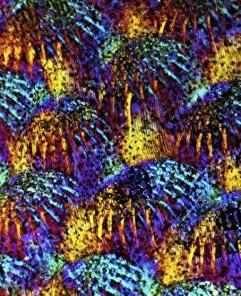
Background imageImages Dated 23rd November 2010: Fish scales, light micrograph

Fish scales, light micrograph
Fish scales. Polarised light micrograph of part of the skin of a fish, showing the placoid protective scales. These scales are found under the epidermis, and are made of bone-like dentine

Background imageImages Dated 23rd November 2010: Butterfly proboscis, light micrograph

Butterfly proboscis, light micrograph
Butterfly proboscis. Light micrograph of the coiled proboscis ( tongue ) of a butterfly. The proboscis of a butterfly consists of two half-tubes which join together in the mid-line to form a single

Background imageImages Dated 23rd November 2010: Strychnine seed tissue, light micrograph

Strychnine seed tissue, light micrograph
Strychnine seed tissue. Light micrograph of a section through a seed of the strychnine plant (Strychnos nux vomica), showing the cellular bridges (plasmodesma)

Background imageImages Dated 23rd November 2010: Tongue papillae, light micrograph

Tongue papillae, light micrograph
Tongue papillae. Light micrograph of a vertical section through circumvallate papillae on a mammalian tongue. A full papilla is at right, with a partially seen one at left

Background imageImages Dated 23rd November 2010: Liverwort leaf tissue, light micrograph

Liverwort leaf tissue, light micrograph
Liverwort leaf tissue. Light micrograph of a leaf from a leafy liverwort (Lophocolea cuspidata). These leaves are extremely thin, consisting of one layer of cells full of chloroplasts

Background imageImages Dated 23rd November 2010: St Ives

St Ives
Colourful fenders on the side of little open Fishing Boats lit by pale afternoon sun light, in the harbour at St Ives on the north coast of Cornwall, on a winters afternoon

Background imageImages Dated 23rd November 2010: Cape Cornwall

Cape Cornwall in Cornwall, is four miles north of Lands End near the town of St Just. looking out into the Atlantic

Background imageImages Dated 23rd November 2010: Working Boat

Working Boat
A working Boat near Lands End Cornwall

Background imageImages Dated 23rd November 2010: Cape Cornwall

Cape Cornwall in Cornwall, is four miles north of Lands End near the town of St Just. looking out into the Atlantic

Background imageImages Dated 23rd November 2010: Botallack Tin Mine

Botallack Tin Mine
The Botallack Tin Mine on the coast of Cornwall near St Just, on a winters day

Background imageImages Dated 23rd November 2010: Cape Cornwall

Cape Cornwall in Cornwall, is four miles north of Lands End near the town of St Just. looking out into the Atlantic


All Professionally Made to Order for Quick Shipping


-

Why Choose Us?

We are a leading provider of Art Prints and Photo Gifts since 2004, working in partnership with a range of Sporting Clubs, Charities, Museums and Picture Libraries. A large share of profits from any of their images will go directly towards supporting that charity or club. Our archive of images is carefully curated to bring you a wide range of subjects, including landscapes, wildlife, architecture, and more. We ship from our partner labs in the UK, USA, EU (Netherlands) and Australia.
+

How do I place an order?

Ordering is quick & easy - Just follow 5 Simple Steps:
  1. Find your image: Use our search box or browse our online photo Collections to find the image you want.

  2. Choose your print format: Select your desired product and add it to your cart.

  3. Enter your details: If you're a returning customer, simply enter your email address and password, and we'll fill in your billing and shipping address details. All personal details are held securely and are fully GDPR compliant. As standard, we remove all Personally Identifiable Information after 12 months.

  4. Pay for your purchase: We use state-of-the-art security for online shopping and do not have access to your card details.

  5. Sit back and relax: We'll email you confirmation of your order and when it's dispatched. Registered customers can also track orders in the 'My Account' area.

+

How do I pay for an order?

You can pay for your order with most of the major credit and debit cards, or PayPal. For added security, major financial institutions process payment details separately and securely on our behalf. We do not have access to your online payment card details. Online payments are preferred however we do take phone orders. For UK customers only we accept cheques issued against a UK bank.
+

Is my data safe?

We take data security very seriously. We do not have access to your full card details and all payments are requested over a fully secure connection. Additionally, we fully comply with current European and GDPR legislation, and automatically remove all personal data after at most 12 months (unless you have an account with us and have signed in recently). We also have a strict opt-in policy and would never sell your personal details. Your data is only used to fulfill your order promptly and efficiently.
+

Is wall art ready to hang?

For quick and easy installation all wall art, including framed prints, canvas prints and metal prints are supplied with a ready to hang solution on the back. Generally, saw tooth hangers are applied as they allow wall art to hang flush against the wall. The serrated edge of the hanger prevents the frame from shifting or tilting when hung.
+

Are the photo prints fade resistant?

Yes, we use archival quality photo paper photographic paper for vivid reproduction Prints are an accurate representations of the original artwork, which is preserved for artistic character and authenticity. We guarantee they match previews shown on our web site