Fraud Blocker Skip to main content

Home > All Images > 2011 > December > 21 Dec 2011

Images Dated 21st December 2011 (page 7)

613 items

Background imageImages Dated 21st December 2011: Country lane with farm buildings

Country lane with farm buildings
A country lane with farm buildings

Background imageImages Dated 21st December 2011: Elephants in the water

Elephants in the water
A group of elephants frolicking in the water

Background imageImages Dated 21st December 2011: Study of a heron in flight

Study of a heron in flight

Background imageImages Dated 21st December 2011: Scene outside a country church

Scene outside a country church, showing people arriving for a service

Background imageImages Dated 21st December 2011: Country scene with farm building

Country scene with farm building
A country scene with a farm building and a grassy field

Background imageImages Dated 21st December 2011: Man carrying an eel trap

Man carrying an eel trap
A man carrying a wickerwork eel trap on his back. More traps can be seen in the background

Background imageImages Dated 21st December 2011: Country lane with farm buildings and fields

Country lane with farm buildings and fields
A pretty country lane with farm buildings, and fields extending into the distance

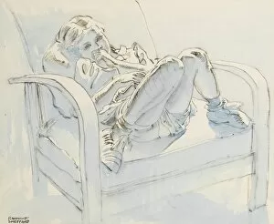
Background imageImages Dated 21st December 2011: Little girl sitting in a chair

Little girl sitting in a chair
Study of a little girl sitting in a chair

Background imageImages Dated 21st December 2011: Duck in flight

Duck in flight
Illustration of a duck in flight

Background imageImages Dated 21st December 2011: Watercolour study of birds

Watercolour study of birds

Background imageImages Dated 21st December 2011: Rocky landscape with trees and grass

Rocky landscape with trees and grass
A rocky landscape with trees and grass

Background imageImages Dated 21st December 2011: Boy on a horse with wild animals

Boy on a horse with wild animals
Illustration of a boy riding a horse, followed by various wild animals, including a lion, a rhinoceros and elephants

Background imageImages Dated 21st December 2011: Portrait of a young boy

Portrait of a young boy wearing a shirt, school tie and knitted sleeveless jumper

Background imageImages Dated 21st December 2011: Primitive sailing boat

Primitive sailing boat
A primitive sailing boat near an island, probably in the Pacific Ocean

Background imageImages Dated 21st December 2011: Sketch of a country scene

Sketch of a country scene

Background imageImages Dated 21st December 2011: Jerusalem / Mount Olives

Jerusalem / Mount Olives
Jerusalem: the Mount of Olives viewed from Jerusalem Date: 1910

Background imageImages Dated 21st December 2011: Jerusalem / Mount Olives

Jerusalem / Mount Olives
The Mount of Olives

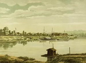
Background imageImages Dated 21st December 2011: Borneo / Sintang 1883

Borneo / Sintang 1883
Borneo: general view of Sintang Date: 1883

Background imageImages Dated 21st December 2011: Jerusalem / Mount Olives

Jerusalem / Mount Olives
Jerusalem: the Mount of Olives, viewed from Mount Zion Date: 1912

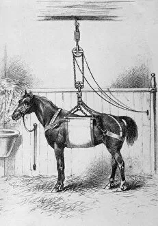
Background imageImages Dated 21st December 2011: Horse in Sling

Horse in Sling
A horse is placed in a sling by the vet, in order that he can examine and treat it more easily. Date: 1886

Background imageImages Dated 21st December 2011: Bob Champion

Bob Champion
Horse Racing - 1993 Imperial Cup - Sandown Park Bob Champion, who famously won the 1981 Grand National on Aldaniti

Background imageImages Dated 21st December 2011: Sharp rush stem, light micrograph

Sharp rush stem, light micrograph
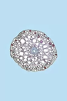
Sharp rush stem. Light micrograph of a section through the stem of a sharp rush (Juncus acutus) plant. This arid-adapted plant (xerophyte) has scattered vascular bundles

Background imageImages Dated 21st December 2011: Acorus calamus rhizome, light micrograph

Acorus calamus rhizome, light micrograph
Acorus calamus rhizome. Light micrograph of a section through a rhizome from a sweet flag (Acorus calamus) plant. The rhizome has an outer layer with scattered collateral vascular bundles

Background imageImages Dated 21st December 2011: Maize stem, light micrograph

Maize stem, light micrograph
Maize stem. Light micrograph of a section through the stem of a maize (corn, Zea mays) plant. The outer epidermis has a hypodermis of thick-walled cells of sclerenchyma (red)

Background imageImages Dated 21st December 2011: Ammophila arenaria leaf, light micrograph

Ammophila arenaria leaf, light micrograph
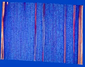
Ammophila arenaria leaf. Polarised light micrograph of a section through a marram grass (Ammophila arenaria) leaf, showing the characteristics that help reduce water loss

Background imageImages Dated 21st December 2011: Lily flower bud, light micrograph

Lily flower bud, light micrograph
Lily flower bud. Light micrograph of a section through a pollen sac from an anther of a lily (Lilium sp.) flower bud, showing mature pollen grains (dark)

Background imageImages Dated 21st December 2011: Ammophila arenaria leaf, light micrograph

Ammophila arenaria leaf, light micrograph
Ammophila arenaria leaf. Polarised light micrograph of a section through a marram grass (Ammophila arenaria) leaf, showing the characteristics that help reduce water loss

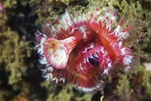
Background imageImages Dated 21st December 2011: Polychaete marine worm, Japan

Polychaete marine worm, Japan
Polychaete marine worm (Serpula uschakovi), Japan

Background imageImages Dated 21st December 2011: Dendrobium orchid root, light micrograph

Dendrobium orchid root, light micrograph
Dendrobium orchid root. Light micrograph of a section through an aerial root from a Dendrobium sp. orchid. The outer tissue (velamen radicum, not shown) is composed of hexagonal cells

Background imageImages Dated 21st December 2011: Starfish preying in sea urchin

Starfish preying in sea urchin
Starfish (Lysastrosoma anthosticta) preying on a sea urchin (Strongylocentrotus nudus) Japan

Background imageImages Dated 21st December 2011: Dendrobium orchid root, light micrograph

Dendrobium orchid root, light micrograph
Dendrobium orchid root. Light micrograph of a section through an aerial root from a Dendrobium sp. orchid. The outer tissue (velamen radicum, grey) is composed of hexagonal cells

Background imageImages Dated 21st December 2011: Maize stem, light micrograph

Maize stem, light micrograph
Maize stem. Polarised light micrograph of a section through the stem of a maize (corn, Zea mays) plant, showing six long vascular bundles surrounded by the box-shaped cells of parenchyma tissue

Background imageImages Dated 21st December 2011: Ammophila arenaria leaf, light micrograph

Ammophila arenaria leaf, light micrograph
Ammophila arenaria leaf. Light micrograph of a section through a marram grass (Ammophila arenaria) leaf, showing the characteristics that help reduce water loss

Background imageImages Dated 21st December 2011: Smilax root, light micrograph

Smilax root, light micrograph
Smilax root. Light micrograph of a section through a root from a China root (Smilax china) plant. The outer epidermis has old root hairs (pink). Below the epidermis are thick-walled cork cells (red)

Background imageImages Dated 21st December 2011: Canadian pondweed leaf, light micrograph

Canadian pondweed leaf, light micrograph
Canadian pondweed leaf. Light micrograph of a section through a leaf from a Canadian pondweed (Elodea canadiensis) plant. The centre of the stem has a vascular system (blue)

Background imageImages Dated 21st December 2011: Ammophila arenaria leaf, light micrograph

Ammophila arenaria leaf, light micrograph
Ammophila arenaria leaf. Light micrograph of a section through a marram grass (Ammophila arenaria) leaf, showing the characteristics that help reduce water loss

Background imageImages Dated 21st December 2011: Lily flower ovary, light micrograph

Lily flower ovary, light micrograph
Lily flower ovary. Light micrograph of a section through an ovule (ovary) from a lily (Lilium sp.) flower, showing the embryo sac (oval, centre left)

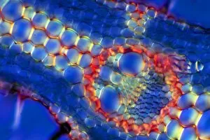
Background imageImages Dated 21st December 2011: Dracaena draco stem, light micrograph

Dracaena draco stem, light micrograph
Dracaena draco stem. Light micrograph of a section through the stem of a young dragon tree (Dracaena draco). Shown here is an outer ring of vascular bundles, containing phloem (blue) and xylem (red)

Background imageImages Dated 21st December 2011: Orchid pseudo bulb, light micrograph

Orchid pseudo bulb, light micrograph
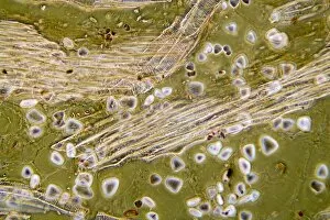
Orchid pseudo bulb. Light micrograph of a section through the bulb of a Phaius grandifolia orchid, showing large starch grains (triangular)

Background imageImages Dated 21st December 2011: Acorus calamus rhizome, light micrograph

Acorus calamus rhizome, light micrograph
Acorus calamus rhizome. Light micrograph of a section through a rhizome from a sweet flag (Acorus calamus) plant. The rhizome has an outer layer with scattered collateral vascular bundles

Background imageImages Dated 21st December 2011: Wheat leaf, light micrograph

Wheat leaf, light micrograph
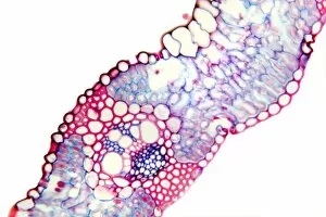
Wheat leaf. Light micrograph of a section through a leaf from a common wheat (Triticum aestivum) plant. The vascular bundle (lower centre), or vein

Background imageImages Dated 21st December 2011: Wheat leaf, light micrograph

Wheat leaf, light micrograph
Wheat leaf. Light micrograph of a section through a leaf from a common wheat (Triticum aestivum) plant. The vascular bundle (centre to upper centre), or vein

Background imageImages Dated 21st December 2011: Common rush stem, light micrograph

Common rush stem, light micrograph
Common rush stem. Light micrograph of a section through the stem of a common rush (Juncus conglomeratus) plant, showing stellate cells

Background imageImages Dated 21st December 2011: Canadian pondweed leaf, light micrograph

Canadian pondweed leaf, light micrograph
Canadian pondweed leaf. Light micrograph of a section through a leaf from a Canadian pondweed (Elodea canadiensis) plant. Magnification: x5 when printed 10 centimetres wide

Background imageImages Dated 21st December 2011: Dendrobium orchid root, light micrograph

Dendrobium orchid root, light micrograph
Dendrobium orchid root. Light micrograph of a section through an aerial root from a Dendrobium sp. orchid. The outer tissue (velamen radicum, grey) is composed of hexagonal cells

Background imageImages Dated 21st December 2011: Common rush stem, light micrograph

Common rush stem, light micrograph
Common rush stem. Light micrograph of a section through the stem of a common rush (Juncus conglomeratus) plant, showing stellate cells

Background imageImages Dated 21st December 2011: Hydrozoan medusa

Hydrozoan medusa (Gonionemus vertens), Japan

Background imageImages Dated 21st December 2011: Lily flower bud, light micrograph

Lily flower bud, light micrograph
Lily flower bud. Light micrograph of a section through a young flower bud from a lily (Lilium sp.), showing pollen cells (circles) dividing within the anthers (inner ring)


All Professionally Made to Order for Quick Shipping


-

Why Choose Us?

We are a leading provider of Art Prints and Photo Gifts since 2004, working in partnership with a range of Sporting Clubs, Charities, Museums and Picture Libraries. A large share of profits from any of their images will go directly towards supporting that charity or club. Our archive of images is carefully curated to bring you a wide range of subjects, including landscapes, wildlife, architecture, and more. We ship from our partner labs in the UK, USA, EU (Netherlands) and Australia.
+

How do I place an order?

Ordering is quick & easy - Just follow 5 Simple Steps:
  1. Find your image: Use our search box or browse our online photo Collections to find the image you want.

  2. Choose your print format: Select your desired product and add it to your cart.

  3. Enter your details: If you're a returning customer, simply enter your email address and password, and we'll fill in your billing and shipping address details. All personal details are held securely and are fully GDPR compliant. As standard, we remove all Personally Identifiable Information after 12 months.

  4. Pay for your purchase: We use state-of-the-art security for online shopping and do not have access to your card details.

  5. Sit back and relax: We'll email you confirmation of your order and when it's dispatched. Registered customers can also track orders in the 'My Account' area.

+

How do I pay for an order?

You can pay for your order with most of the major credit and debit cards, or PayPal. For added security, major financial institutions process payment details separately and securely on our behalf. We do not have access to your online payment card details. Online payments are preferred however we do take phone orders. For UK customers only we accept cheques issued against a UK bank.
+

Is my data safe?

We take data security very seriously. We do not have access to your full card details and all payments are requested over a fully secure connection. Additionally, we fully comply with current European and GDPR legislation, and automatically remove all personal data after at most 12 months (unless you have an account with us and have signed in recently). We also have a strict opt-in policy and would never sell your personal details. Your data is only used to fulfill your order promptly and efficiently.
+

Is wall art ready to hang?

For quick and easy installation all wall art, including framed prints, canvas prints and metal prints are supplied with a ready to hang solution on the back. Generally, saw tooth hangers are applied as they allow wall art to hang flush against the wall. The serrated edge of the hanger prevents the frame from shifting or tilting when hung.
+

Are the photo prints fade resistant?

Yes, we use archival quality photo paper photographic paper for vivid reproduction Prints are an accurate representations of the original artwork, which is preserved for artistic character and authenticity. We guarantee they match previews shown on our web site