Home > Fine Art Storehouse > Science Inspired Art
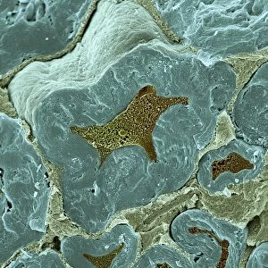
Xylem, LM. Xylem tissue. Light micrograph (LM) of a section through sunflower
 of a section through sunflower")
![]()
Wall Art and Photo Gifts from Fine Art Storehouse
Xylem, LM. Xylem tissue. Light micrograph (LM) of a section through sunflower
Xylem tissue. Light micrograph (LM) of a section through sunflower(helianthus annuus) tissue showing spiral tracheids, a type of xylem. Tracheids are long tubular cells with lignin, a material that provides support, in the cell walls. Spiral thickening of the cells can be seen. Tracheids conduct water from the roots of a plant along the stems to the leaves. Magnification: x210 when printed 10cm wide
Unleash your creativity and transform your space into a visual masterpiece!
STEVE GSCHMEISSNER/SCIENCE PHOTO
Media ID 19527365
© Science Photo Library
Biologica Biology Horizontal Image Scientific
FEATURES IN THESE COLLECTIONS
> Fine Art Storehouse
> Science Inspired Art
> SEM (Scanning Electron Microscope)
> Fine Art Storehouse
> Science Inspired Art
EDITORS COMMENTS
This print captures the intricate beauty of xylem tissue in a sunflower. The light micrograph reveals a section through the tissue, showcasing spiral tracheids, a specific type of xylem cells. These long tubular cells play a vital role in plant physiology by conducting water from the roots to the leaves. The image showcases the remarkable structure and functionality of these tracheids. Lignin, which provides support and strength to plants, is clearly visible in their cell walls. Additionally, one can observe the fascinating spiral thickening pattern within these cells. Taken at a magnification of x210 and printed 10cm wide, this horizontal photograph allows us to appreciate nature's microscopic wonders on a larger scale. It invites viewers into an intriguing world where science meets art. With its scientific focus and meticulous detail, this image appeals not only to biologists but also to those with an appreciation for natural beauty. Its composition highlights both precision and elegance while offering insight into the inner workings of plant life. Whether displayed in educational institutions or as part of personal collections, this stunning print serves as a reminder that even at microscopic levels, nature never ceases to amaze us with its complexity and aesthetic appeal.
MADE IN AUSTRALIA
Safe Shipping with 30 Day Money Back Guarantee
FREE PERSONALISATION*
We are proud to offer a range of customisation features including Personalised Captions, Color Filters and Picture Zoom Tools
SECURE PAYMENTS
We happily accept a wide range of payment options so you can pay for the things you need in the way that is most convenient for you
* Options may vary by product and licensing agreement. Zoomed Pictures can be adjusted in the Cart.