Home > Popular Themes > Human Body
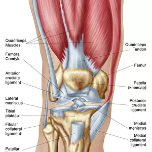
Bovie used to cut through retincaculum, and clean up femur of Displaced patellar knee

![]()
Wall Art and Photo Gifts from Fine Art Storehouse
Bovie used to cut through retincaculum, and clean up femur of Displaced patellar knee
Detail, Anatomy, Diagram, Muscle, Illustration, Bone, Cutting, Equipment, Injury, Medical, Tool, Biology, Femur, Surgery, Knee, Torn, Fractured, Meniscus, Cut Out, White Background, Vertical, Color Image, Front View, Close Up, Healthcare And Medicine, The Human Body, Human Anatomy, No People, Part Of, Human Bone, Surgical Procedure, Cartilage, Human Body Part, Human Knee, patella, fibula, inflammation, tibia, ligament, inflamed, Anterior knee joint, patellar ligament, quadraceps femoris muscles, cruciate, knee bone, Bovie, biomedical illustration, human joint, joint - body part, quadraceps femoris, retinaculum, Illustrations & Artwork, 667600605
Unleash your creativity and transform your space into a visual masterpiece!
Lauren Shavell / Design Pics
Media ID 20655501
Anatomy Biology Biomedical Illustration Bone Cartilage Close Up Cutting Detail Diagram Equipment Femur Fibula Healthcare And Medicine Human Anatomy Human Bone Human Joint Human Knee Inflamed Inflammation Injury Joint Body Part Knee Ligament Medical Muscle Surgery The Human Body Tibia Tool Torn Fractured Human Body Part Meniscus Patella Patellar Ligament Surgical Procedure
FEATURES IN THESE COLLECTIONS
> Fine Art Storehouse
> Science Inspired Art
> Illustrations & Artwork
> Fine Art Storehouse
> Science Inspired Art
EDITORS COMMENTS
This print by Lauren Shavell showcases the intricate details of a bovie tool being used to cut through the retincaculum and clean up the femur of a displaced patellar knee. The image, set against a pristine white background, provides a close-up view of this medical procedure, highlighting the anatomy and diagram of the knee. The vibrant colors and front view perspective draw attention to every aspect of this surgical process. It is evident that healthcare and medicine play vital roles in our lives as we witness the complexity involved in treating injuries such as torn or fractured knees. The illustration not only focuses on bones but also emphasizes muscles, ligaments, cartilage, and other components that make up the human knee joint. The inflamed tissues surrounding the area further emphasize how crucial it is to address these issues with precision. Through this artwork, viewers gain insight into both biology and surgery while appreciating the intricacies of our own bodies. This biomedical illustration serves as an educational tool for students studying human anatomy or individuals seeking knowledge about specific medical procedures. Lauren Shavell's attention to detail captures every element necessary for understanding this particular surgical intervention. Without any people present in the image itself, it allows us to focus solely on this part of our body that often goes unnoticed until injury strikes.
MADE IN AUSTRALIA
Safe Shipping with 30 Day Money Back Guarantee
FREE PERSONALISATION*
We are proud to offer a range of customisation features including Personalised Captions, Color Filters and Picture Zoom Tools
SECURE PAYMENTS
We happily accept a wide range of payment options so you can pay for the things you need in the way that is most convenient for you
* Options may vary by product and licensing agreement. Zoomed Pictures can be adjusted in the Cart.