
Abnormal Collection (page 3)
"Unveiling the Abnormal: A Glimpse into Extraordinary Phenomena" Gas pillars in the Eagle Nebula: Witness the mesmerizing dance of celestial gas pillars


All Professionally Made to Order for Quick Shipping
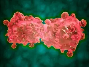
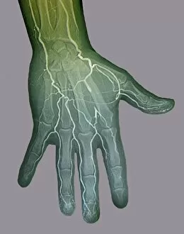
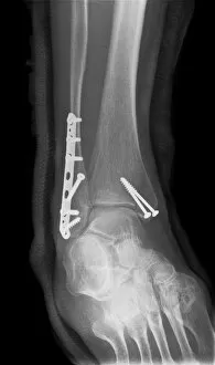
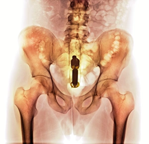
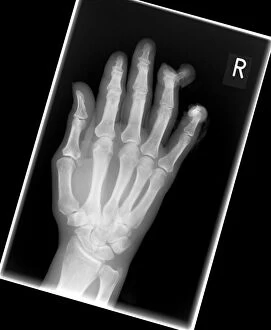
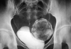
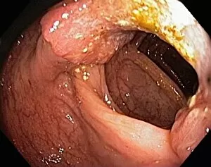
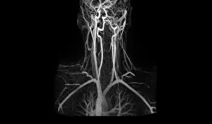
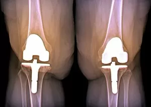
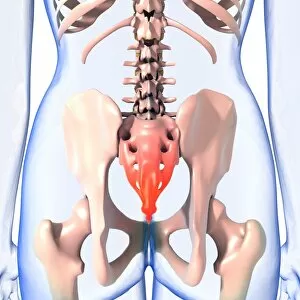
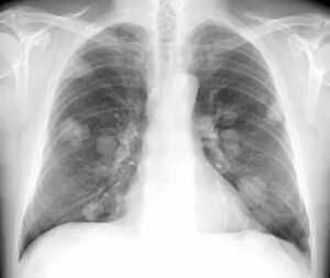
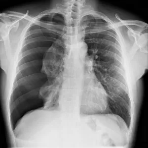
"Unveiling the Abnormal: A Glimpse into Extraordinary Phenomena" Gas pillars in the Eagle Nebula: Witness the mesmerizing dance of celestial gas pillars, defying gravity and painting a surreal picture in the depths of space. Apollo 11 astronaut footprint on Moon: Behold an iconic abnormality, a single footprint etched by humanity's giant leap onto another world, forever marking our presence among the stars. Broken wrist bone, X-ray C017 / 7187: Delve into the intricate realm of human anatomy as we explore an abnormality captured within an X-ray image – a fractured wrist bone telling tales of resilience and healing. Solar prominence: Marvel at nature's fiery spectacle as solar prominences erupt from our nearest star, showcasing their abnormal beauty against the backdrop of infinite cosmic expanse. DAF 95 XF wide: Discover automotive innovation with this extraordinary truck model boasting exceptional width – its bold design challenges conventional norms to redefine road transport. HeLa cells, light micrograph C017 / 8299: Peer through a microscope lens to witness HeLa cells – immortalized yet controversial entities that have revolutionized medical research while raising ethical questions about their origin. Image of Jupiter taken with Hubble Telescope: Explore Jupiter's enigmatic atmosphere through an extraordinary image captured by Hubble Telescope; revealing swirling storms and vibrant bands that defy expectations. Knee pain, conceptual artwork: Dive into thought-provoking artistry depicting knee pain – a visual representation inviting contemplation on physical discomforts experienced by many but often hidden beneath societal masks. Joint pain, conceptual artwork: Engage your senses with evocative imagery portraying joint pain; transcending boundaries between art and reality to shed light on invisible struggles faced by individuals worldwide. Total hip replacement, X-ray.