Fraud Blocker Skip to main content

Abnormal Collection (page 4)

"Unveiling the Abnormal: A Glimpse into Extraordinary Phenomena" Gas pillars in the Eagle Nebula: Witness the mesmerizing dance of celestial gas pillars

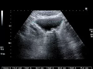
Background imageAbnormal Collection: Bladder stones, ultrasound scan C017 / 7974

Bladder stones, ultrasound scan C017 / 7974
Bladder stones. Ultrasound scan of the bladder of a 58 year old male showing bladder stones (bright white). Bladder stones (cystoliths)

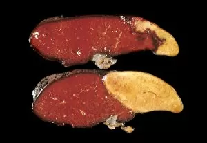
Background imageAbnormal Collection: Splenic infarct C015 / 6220

Splenic infarct C015 / 6220
Splenic infarct. Gross specimen of a sectioned spleen showing areas of dead tissue (pale). The tissue death results from a loss of blood flow to the area, known as an infarction

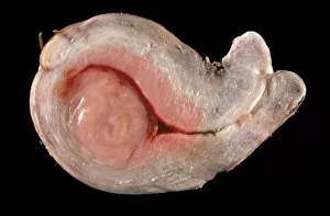
Background imageAbnormal Collection: Uterine fibroid C015 / 6407

Uterine fibroid C015 / 6407
Uterine fibroid. Gross specimen of a sectioned uterine fibroid (left). A fibroid is a fibrous benign tumour originating from muscular tissue

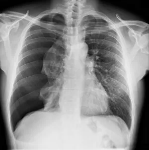
Background imageAbnormal Collection: Pneumothorax, X-ray C017 / 7812

Pneumothorax, X-ray C017 / 7812
Pneumothorax. X-ray of the chest of a patient with a pneumothorax, or collapsed lung. The right lung (left on X-ray) has collapsed due to a build-up of air (solid grey)

Background imageAbnormal Collection: Hydropneumothorax, X-ray C017 / 7810

Hydropneumothorax, X-ray C017 / 7810
Hydropneumothorax. X-ray of the chest of a female patient with a hydropneumothorax of the right lung (seen at left). This is a condition where air and fluid accumulate in the spaces around the lungs

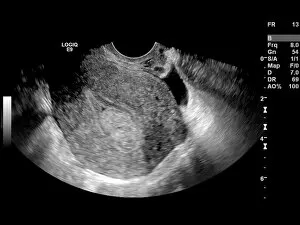
Background imageAbnormal Collection: Endometrial polyp, ultrasound scan C017 / 7800

Endometrial polyp, ultrasound scan C017 / 7800
Endometrial polyp. Ultrasound scan of the uterus (womb) of a 42 year old female patient with a polyp (lower centre) in the endometrium, the lining of the womb

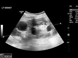
Background imageAbnormal Collection: Polycystic kidney, ultrasound scan C017 / 7746

Polycystic kidney, ultrasound scan C017 / 7746
Polycystic kidney. Ultrasound scan of the kidney of a 42 year old female showing numerous fluid-filled cysts (dark areas)
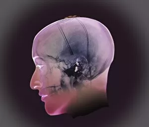
Background imageAbnormal Collection: Deep brain stimulation, X-ray

Deep brain stimulation, X-ray
Image Xrays Crane (Sur Projection 3D Modèle) En Incidence De Profil Chez Une Patiente De 54 Ans, Parkinsonienne Traitee Sous Protocole De Neurochirurgie; Visualisation Des Electrodes De

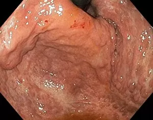
Background imageAbnormal Collection: Severe gastritis, endoscope view C016 / 3898

Severe gastritis, endoscope view C016 / 3898
Severe gastritis, endoscope view. Gastritis is inflammation of the stomach lining. It causes pain on eating or drinking, and may cause nausea and vomiting
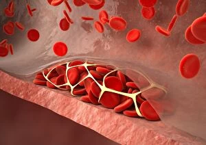
Background imageAbnormal Collection: Thrombosed blood vessel, artwork C013 / 4649

Thrombosed blood vessel, artwork C013 / 4649
Thrombosed blood vessel. Computer artwork of a blood clot (thrombus) formed on the internal wall of a blood vessel

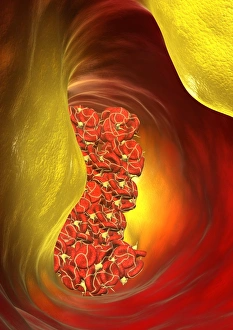
Background imageAbnormal Collection: Blocked artery, computer artwork

Blocked artery, computer artwork
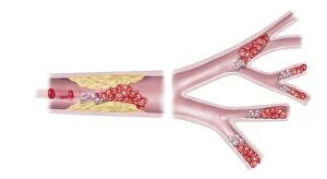
Blocked artery. Computer artwork of a clot of blood cells (thrombus, red) in an artery affected by atherosclerosis. This is a narrowing of an artery due to fatty deposits (yellow) on its inner walls

Background imageAbnormal Collection: Aortic dissection, 3D CT scan

Aortic dissection, 3D CT scan
Aortic dissection. 3D computed tomography (CT) scan of a 67-year-old man with an aortic dissection. The aorta, the bodys main artery, is running vertically down centre (red)

Background imageAbnormal Collection: Dislocated neck bones, X-ray

Dislocated neck bones, X-ray
X-ray (side view) of the neck in a 57 year old patient showing dislocated vertebral bones (C5-C6) after a car accident

Background imageAbnormal Collection: Eating disorder, conceptual artwork

Eating disorder, conceptual artwork

Background imageAbnormal Collection: Leucistic european robin

Leucistic european robin (Erithracus rubecula) perched on a rock. Leucism is a condition characterized by reduced pigmentation in animals and humans

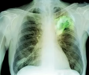
Background imageAbnormal Collection: Lung abscess, X-ray

Lung abscess, X-ray
Lung abscess. Coloured X-ray showing an abscess (dense coloured area, upper right) in the upper lobe of a patients left lung (right). An abscess is an accumulation of puss within a cavity in tissue

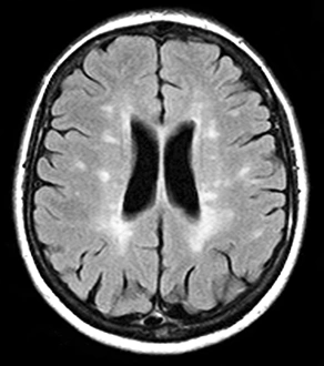
Background imageAbnormal Collection: Multiple sclerosis, MRI scan

Multiple sclerosis, MRI scan

Background imageAbnormal Collection: Hairy-Faced Women

Hairy-Faced Women
Women with abnormal growths of hair on their faces

Background imageAbnormal Collection: Hair on Back

Hair on Back
A lady with an abnormal growth of hair on her back Date: late 19th century

Background imageAbnormal Collection: Hair in Small of Back

Hair in Small of Back
Two people with abnormal growths of hair in the small of the back Date: late 19th century

Background imageAbnormal Collection: Heart with arteries showing cholesterol in one and plaque in the other

Heart with arteries showing cholesterol in one and plaque in the other
Heart with arteries showing cholesterol in one artery and atherosclerotic plaque in the other

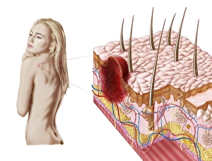
Background imageAbnormal Collection: Illustration of an atypical growth on the skin that could be a sign of skin cancer

Illustration of an atypical growth on the skin that could be a sign of skin cancer
Illustration of an atypical growth on the skin that could be a warning sign of skin cancer. Sudden change in the appearance, shape, color, size

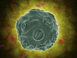
Background imageAbnormal Collection: Conceptual image of the Human Papilloma Virus

Conceptual image of the Human Papilloma Virus (HPV). HPV is a virus from the papillomavirus family that affects human skin and the moist membranes that line the body

Background imageAbnormal Collection: Blood flow through a relaxed artery verus an artery in spasm

Blood flow through a relaxed artery verus an artery in spasm

Background imageAbnormal Collection: Trachea cross-section showing normal and asthmatic bronchiole

Trachea cross-section showing normal and asthmatic bronchiole
Trachea cross-section showing comparison of normal and asthmatic bronchiole

Background imageAbnormal Collection: Development of fistula

Development of fistula

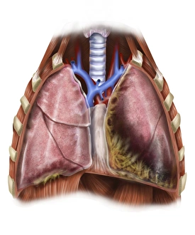
Background imageAbnormal Collection: Artist depiction of mesothelioma in the lungs / abdominal cavity

Artist depiction of mesothelioma in the lungs / abdominal cavity
Artist depiction of mesothelioma in the lungs/abdominal cavity

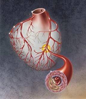
Background imageAbnormal Collection: Arteries on heart showing atherosclerotic plaque in an artery

Arteries on heart showing atherosclerotic plaque in an artery

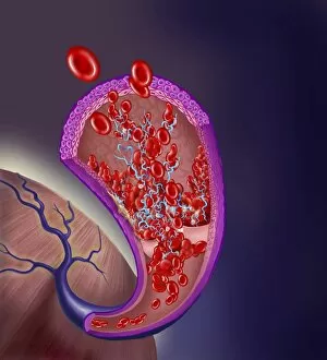
Background imageAbnormal Collection: Microscopic view of blood clotting inside the artery

Microscopic view of blood clotting inside the artery

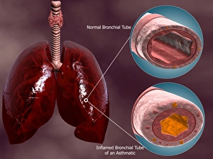
Background imageAbnormal Collection: Trachea cross-section with lungs showing normal and asthmatic bronchiole

Trachea cross-section with lungs showing normal and asthmatic bronchiole
Trachea cross-section with lungs, showing comparison of normal and asthmatic bronchiole

Background imageAbnormal Collection: Good and bad cholesterol found in blood stream

Good and bad cholesterol found in blood stream

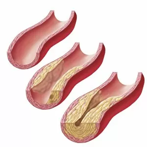
Background imageAbnormal Collection: Build-up of atherosclerotic plaque in an artery

Build-up of atherosclerotic plaque in an artery

Background imageAbnormal Collection: Large eroded plaque on the leg with areas of crust

Large eroded plaque on the leg with areas of crust
Metastatic Melanoma. Large eroded plaque on the leg with areas of crust. Scattered eroded dermal nodules are also present

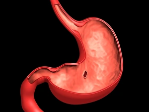
Background imageAbnormal Collection: Conceptual image of peptic ulcer in human stomach

Conceptual image of peptic ulcer in human stomach

Background imageAbnormal Collection: Melanoma on a patients skin

Melanoma on a patients skin

Background imageAbnormal Collection: Acute coronary syndrome - microvascular obstruction

Acute coronary syndrome - microvascular obstruction

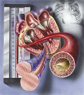
Background imageAbnormal Collection: Interior view of heart with detail of muscle cells and atherosclerotic artery

Interior view of heart with detail of muscle cells and atherosclerotic artery. Chest x-ray in background

Background imageAbnormal Collection: Thrombus forming on valve within vein

Thrombus forming on valve within vein

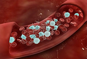
Background imageAbnormal Collection: Blood vessel with platelets, white blood cells and red blood cells

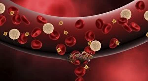
Blood vessel with platelets, white blood cells and red blood cells

Background imageAbnormal Collection: Normal artery compared to plaque and thrombus formation in artery

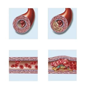
Normal artery compared to plaque and thrombus formation in artery

Background imageAbnormal Collection: Multiphoton fluorescence image of HeLa cells

Multiphoton fluorescence image of HeLa cells with cytoskeletal microtubules (magenta) and DNA (cyan)

Background imageAbnormal Collection: Comparison of normal heart versus heart with a patent foramen ovale

Comparison of normal heart versus heart with a patent foramen ovale

Background imageAbnormal Collection: Comparison of different sized kidney stones in the human kidney

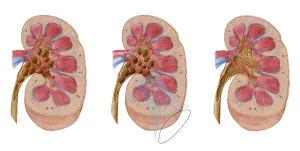
Comparison of different sized kidney stones in the human kidney. The middle image shows the use of extracorporeal shock wave lithotripsy to break up the stones

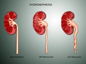
Background imageAbnormal Collection: Hydronephrosis condition of the kidney

Hydronephrosis condition of the kidney
Hydronephrosis is a condition that typically occurs when one kidney becomes swollen due to the failure of normal drainage of urine from the kidney to the bladder

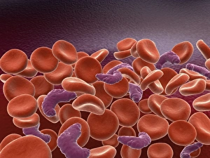
Background imageAbnormal Collection: Conceptual image of sickle cell anemia with red blood cells

Conceptual image of sickle cell anemia with red blood cells

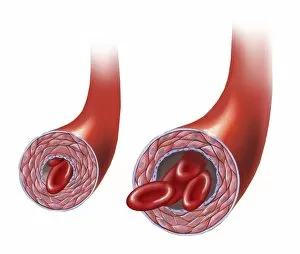
Background imageAbnormal Collection: Normal artery versus artery in spasm

Normal artery versus artery in spasm

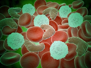
Background imageAbnormal Collection: Sickle cell anemia with red blood cells and white bood cells

Sickle cell anemia with red blood cells and white bood cells
Conceptual image of sickle cell anemia with red blood cells and white bood cells


All Professionally Made to Order for Quick Shipping

"Unveiling the Abnormal: A Glimpse into Extraordinary Phenomena" Gas pillars in the Eagle Nebula: Witness the mesmerizing dance of celestial gas pillars, defying gravity and painting a surreal picture in the depths of space. Apollo 11 astronaut footprint on Moon: Behold an iconic abnormality, a single footprint etched by humanity's giant leap onto another world, forever marking our presence among the stars. Broken wrist bone, X-ray C017 / 7187: Delve into the intricate realm of human anatomy as we explore an abnormality captured within an X-ray image – a fractured wrist bone telling tales of resilience and healing. Solar prominence: Marvel at nature's fiery spectacle as solar prominences erupt from our nearest star, showcasing their abnormal beauty against the backdrop of infinite cosmic expanse. DAF 95 XF wide: Discover automotive innovation with this extraordinary truck model boasting exceptional width – its bold design challenges conventional norms to redefine road transport. HeLa cells, light micrograph C017 / 8299: Peer through a microscope lens to witness HeLa cells – immortalized yet controversial entities that have revolutionized medical research while raising ethical questions about their origin. Image of Jupiter taken with Hubble Telescope: Explore Jupiter's enigmatic atmosphere through an extraordinary image captured by Hubble Telescope; revealing swirling storms and vibrant bands that defy expectations. Knee pain, conceptual artwork: Dive into thought-provoking artistry depicting knee pain – a visual representation inviting contemplation on physical discomforts experienced by many but often hidden beneath societal masks. Joint pain, conceptual artwork: Engage your senses with evocative imagery portraying joint pain; transcending boundaries between art and reality to shed light on invisible struggles faced by individuals worldwide. Total hip replacement, X-ray.