
Acetabulum Collection
The acetabulum, a crucial component of the hip joint, plays a vital role in our mobility and stability


All Professionally Made to Order for Quick Shipping
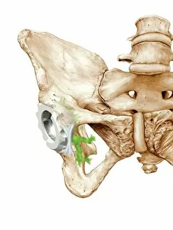
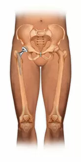
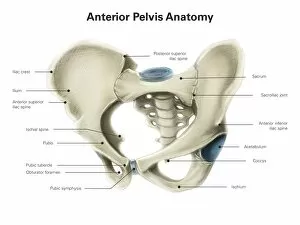
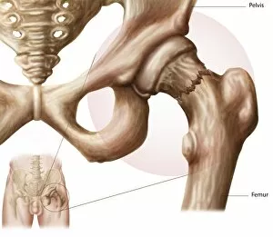
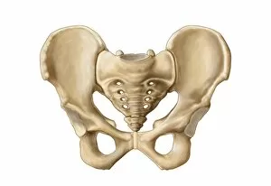
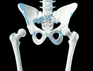
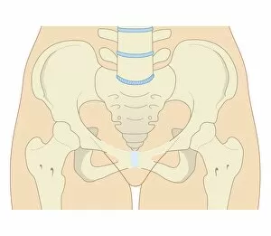
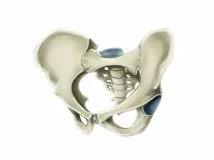
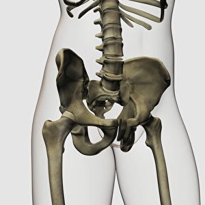
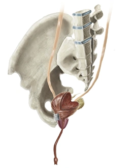
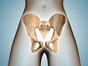
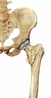
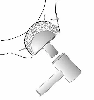
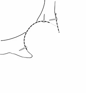
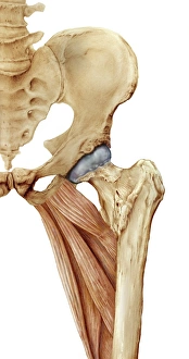
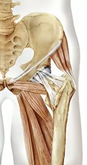
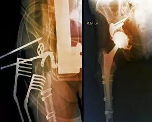
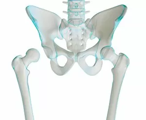
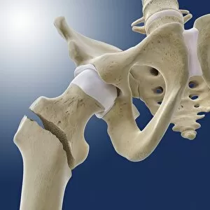
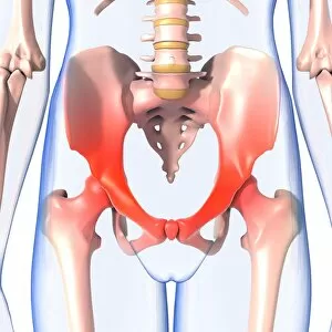
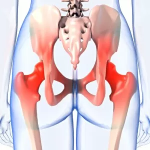
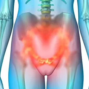
The acetabulum, a crucial component of the hip joint, plays a vital role in our mobility and stability. This concave socket is responsible for connecting the femur to the pelvis, allowing for smooth movement and weight-bearing capabilities. One significant application of the acetabulum is in hip replacement surgeries. When damage or degeneration occurs due to conditions like osteoarthritis or injury, this procedure replaces the damaged joint with an artificial one. The success of such surgeries depends on accurately recreating the shape and function of the acetabulum. Artwork depicting a front view of a body showing a total hip replacement showcases how surgeons meticulously restore functionality by replacing both sides of the joint – including the acetabulum – with prosthetic components. In nature, harbor seals (Phoca vitulina) also rely on their strong hips supported by healthy acetabula to navigate through water effortlessly. These graceful creatures showcase remarkable agility as they glide through their marine habitats. Interestingly, green algae can sometimes find its way into human joints, including the acetabulum. While this may sound unusual, it highlights how intricate our bodies are and how various factors can impact our health. However, not all ailments related to this essential structure involve external elements like algae; some occur internally due to compromised blood supply known as avascular necrosis hip. An illustration demonstrates this condition where insufficient blood flow leads to bone tissue death within the hip joint area. To better understand its anatomy and location within our bodies, we can explore images showcasing anterior views of human pelvises with labeled structures such as bones and organs surrounding it. Moreover, biomedical illustrations provide cross-sectional views that reveal detailed insights into female pelvic bones' three-dimensional structure while highlighting adjacent reproductive systems' complexity. Studying these visuals helps us appreciate just how interconnected our bodies are – from understanding fractures that affect proper functioning to recognizing each component's significance within complex systems like reproduction.