
Alga Collection
"Discovering the Intricate Beauty of Algae: From Ernst Haeckel's Art to 19th-Century Cyanotypes and SEM Images" Immerse yourself in the captivating world of algae


All Professionally Made to Order for Quick Shipping
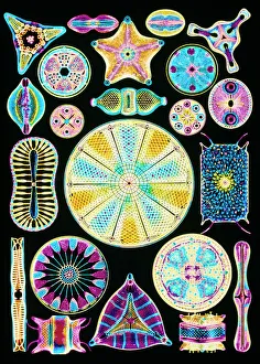
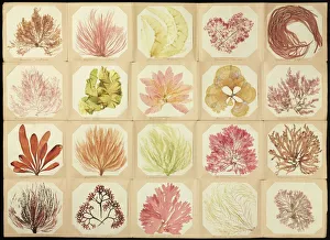
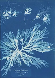
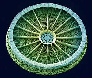
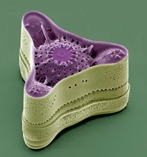
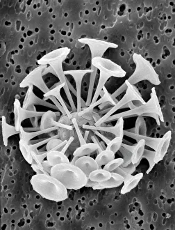
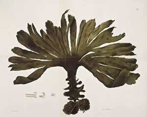
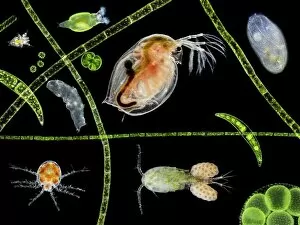
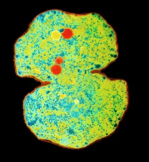
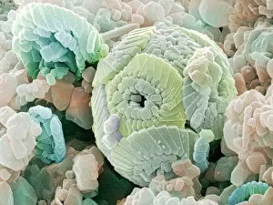
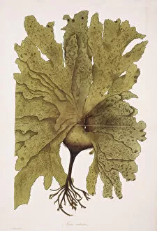
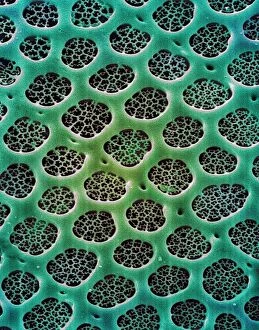
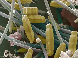
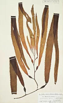
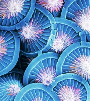
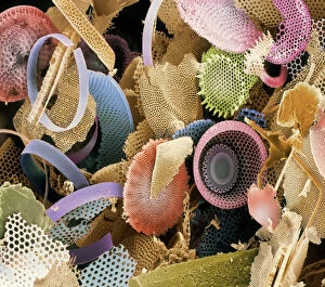
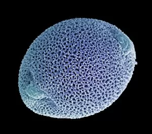
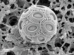
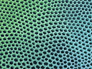
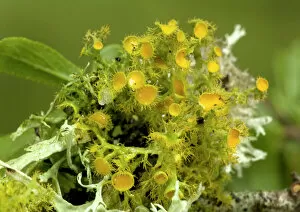
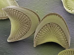
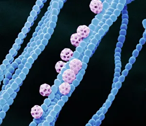
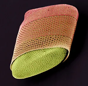
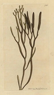
"Discovering the Intricate Beauty of Algae: From Ernst Haeckel's Art to 19th-Century Cyanotypes and SEM Images" Immerse yourself in the captivating world of algae, as we delve into its mesmerizing artistry and scientific wonders. Ernst Haeckel, a renowned biologist and artist, showcases the delicate intricacy of diatom algae through his stunning illustrations. These masterpieces reveal the symmetrical beauty hidden within these microscopic organisms. Step back in time with pressed seaweed specimens C016 / 6127 from centuries ago. Preserved meticulously, they offer a glimpse into the diverse forms and colors that algae can take on. The cyanotype technique used during the 19th century captures their essence in ethereal blue hues, adding an enchanting touch to their already fascinating nature. Calcareous phytoplankton observed under scanning electron microscopy (SEM) unravels another layer of algal marvels. Discosphaera tubifera, a coccolithophore species adorned with intricate calcium carbonate plates, showcases nature's architectural genius at work. Dictyota dichotoma emerges as a striking example of brown algae thriving along coastlines worldwide. Its branching structure creates an artistic spectacle underwater while providing essential habitats for marine life. Diatoms continue to amaze us with their geometric patterns when viewed under SEM imaging techniques. Their microscopic beauty is unveiled through meticulous detailing that reveals their remarkable diversity and ecological significance. Venturing further into aquatic realms, Fucus bulbosus or kelp stands tall as one of nature's giants beneath ocean waves. This majestic alga plays a crucial role in coastal ecosystems by offering shelter to numerous marine organisms. Pond life surprises us with its vibrant assortment of microorganisms where diatoms once again steal the show under SEM imaging techniques. Their intricate designs resemble miniature works of art floating amidst water bodies' tranquility.