
Allergen Collection
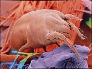
"Allergen: Unseen Culprits Triggering Allergic Reactions" Did you know that tiny creatures like eyelash mites and dust mites can wreak havoc on our immune systems


All Professionally Made to Order for Quick Shipping
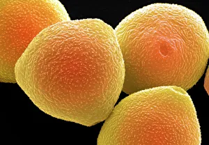
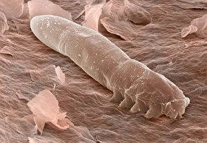
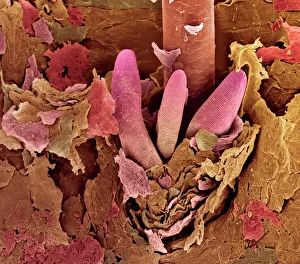
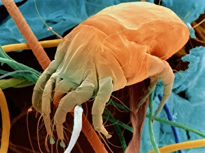
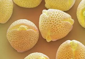
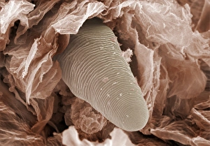
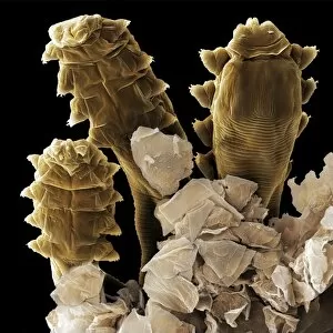
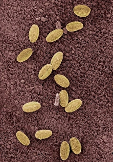
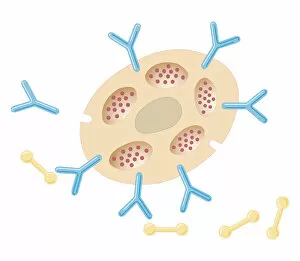
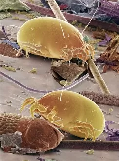
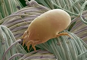
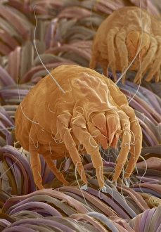
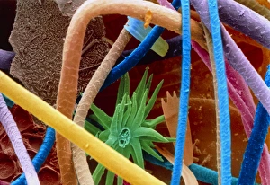
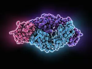
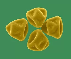
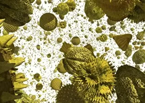
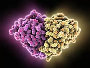
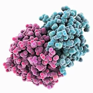
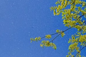
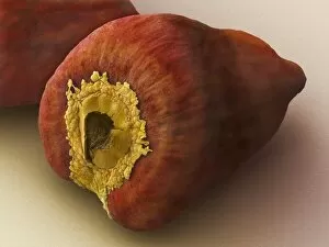
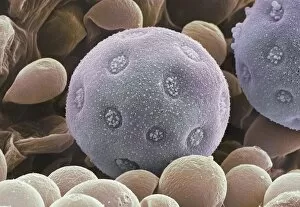
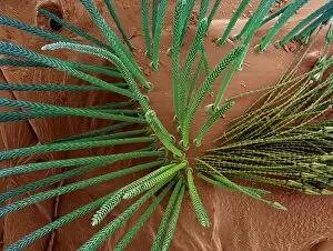
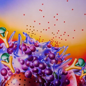
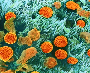
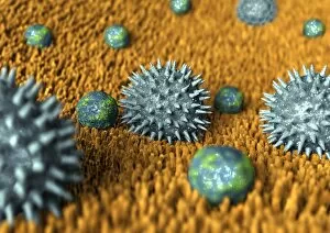
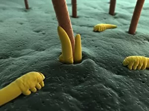
"Allergen: Unseen Culprits Triggering Allergic Reactions" Did you know that tiny creatures like eyelash mites and dust mites can wreak havoc on our immune systems? These microscopic organisms, captured under the lens of a scanning electron microscope (SEM), are often responsible for triggering allergic reactions in many individuals. Take a closer look at the world of allergens. SEM images reveal the intricate details of eyelash mite tails, showcasing their presence on our very own lashes. These minuscule creatures, invisible to the naked eye, can cause itching and irritation for those who are sensitive to them. But it's not just these critters that provoke allergies. Hazel pollen grains and ivy pollen from plants like Hedera helix also play a role in triggering allergic responses. SEM images allow us to observe their unique structures up close, giving insight into how they interact with our bodies. Delving deeper into the microscopic realm, we discover follicle mite heads lurking within hair follicles. These unwelcome guests may contribute to skin irritations and allergies when present in excessive numbers. Allergens don't stop there – they infiltrate even further into our respiratory system. Biomedical illustrations depict allergens making their way into the trachea, causing discomfort and breathing difficulties for allergy sufferers. The battle between allergens and our immune system is illustrated through biomedical cross-sections showing antibodies binding to mast cells after exposure to these troublesome substances. In response, mast cells release histamine – a chemical compound responsible for common allergy symptoms such as sneezing or watery eyes. Dust mites make yet another appearance as notorious allergenic agents. SEM images provide an up-close view of these tiny arachnids that thrive in household environments such as bedding or upholstery materials. Understanding these unseen culprits is crucial in managing allergies effectively.