
Amoeba Collection
"Exploring the Intricate World of Amoebae: From Foraminifer Models to SEM Images" Step into the fascinating world of amoebae


All Professionally Made to Order for Quick Shipping
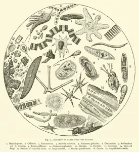
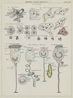
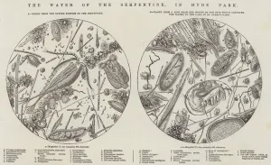
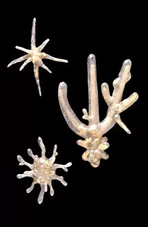
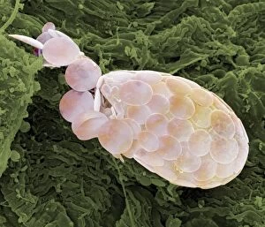
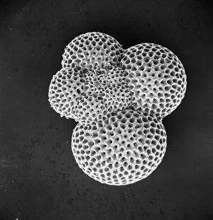
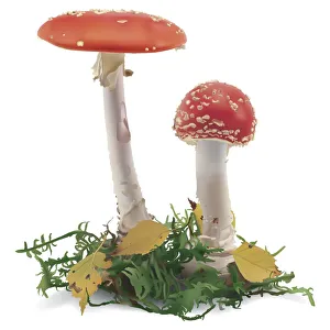
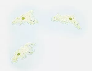
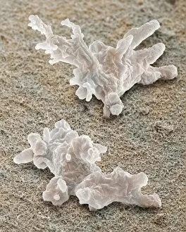
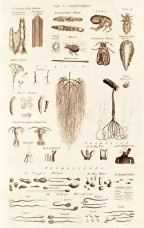
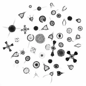
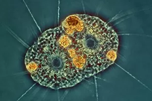
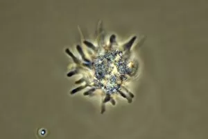
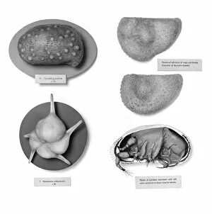
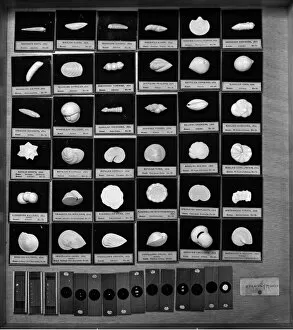
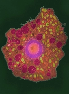
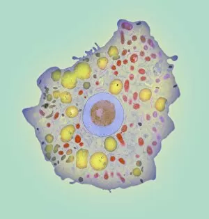
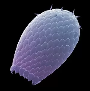
"Exploring the Intricate World of Amoebae: From Foraminifer Models to SEM Images" Step into the fascinating world of amoebae, where intricate forms and microscopic wonders await. The Foraminifer model takes us on a journey through time, showcasing these ancient organisms in all their glory. With its delicate pen strokes and vibrant watercolors, "Imaginary Forms" captures the essence diversity from c. 1939/40. Delving deeper, we encounter the mesmerizing SEM images that reveal the intricate structures proteus. These incredible microorganisms showcase their ever-changing shape and movement, reminding us of nature's boundless creativity. As we explore further, an engraving depicting sediment from London's iconic Thames River captivates our attention. Here lies evidence of amoebae thriving amidst this bustling urban environment – a testament to their adaptability and resilience. Moving on to another captivating lithograph titled "Amoeba, Proteus Animalcule, " we witness a burst of colors that bring these tiny creatures to life. Their translucent bodies seem almost ethereal against the backdrop of scientific precision. Venturing outdoors, an enchanting engraving transports us to Hyde Park's Serpentine waters – home to countless unseen inhabitants like Difflugia pyriformis amoebae. This idyllic scene reminds us that even within bustling cityscapes lie hidden worlds waiting to be discovered. In Picture No. 11014619, we are presented with yet another glimpse into this diverse realm as foraminifers take center stage once again. Their intricate shells tell stories untold as they silently drift through oceans unknown. Stepping away momentarily from amoebae themselves but still within nature's embrace, we encounter Red Aspen Bolete mushrooms standing tall in all their crimson-capped splendor. A reminder that beauty can be found at every scale - whether it be microscopic or towering above the forest floor.