Fraud Blocker Skip to main content

Anatomy Collection (page 19)

"Unveiling the Intricacies of Anatomy: From Sensory Homunculus to Motor Homunculus" Step into the fascinating world of anatomy, where every detail tells a story

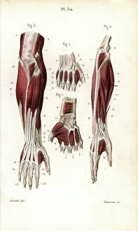
Background imageAnatomy Collection: Human hand - 1866 anatomical illustration showing the muscles (brown) and tendons (white)

Human hand - 1866 anatomical illustration showing the muscles (brown) and tendons (white)
JAB4119450 Human hand - 1866 anatomical illustration showing the muscles (brown) and tendons (white) of the human hand - Region palmar- Anatomy - Fig 1 Cubital anterieur Fig 2 cubital anterieur et

Background imageAnatomy Collection: Leg joints - 1866 anatomical illustration of the muscles (brown), tendons (white) and bones (beige)

Leg joints - 1866 anatomical illustration of the muscles (brown), tendons (white) and bones (beige)
JAB4119440 Leg joints - 1866 anatomical illustration of the muscles (brown), tendons (white) and bones (beige) of the human hip (top) and knee (bottom) joints

Background imageAnatomy Collection: Leg joints - 1866 anatomical illustration of the muscles (brown), tendons (white) and bones (beige)

Leg joints - 1866 anatomical illustration of the muscles (brown), tendons (white) and bones (beige)
JAB4119439 Leg joints - 1866 anatomical illustration of the muscles (brown), tendons (white) and bones (beige) of the human hip (top) and knee (bottom) joints

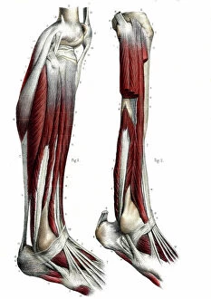
Background imageAnatomy Collection: Leg anatomy - 1866 anatomical illustration showing the muscles (brown)

Leg anatomy - 1866 anatomical illustration showing the muscles (brown), tendons (white) and bones (beige)
JAB4119436 Leg anatomy - 1866 anatomical illustration showing the muscles (brown), tendons (white) and bones (beige) of the human lower leg

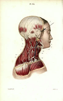
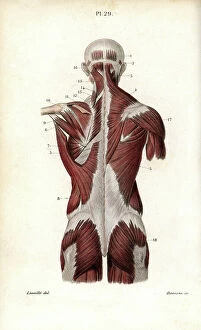
Background imageAnatomy Collection: Chest and neck muscles - 1866 anatomical illustration showing the structure of the muscles (brown)

Chest and neck muscles - 1866 anatomical illustration showing the structure of the muscles (brown) and tendons (white)
JAB4119434 Chest and neck muscles - 1866 anatomical illustration showing the structure of the muscles (brown) and tendons (white) found in the rear of the human head (top) and neck (upper centre)

Background imageAnatomy Collection: Neck muscles - 1866 illustration showing the structure of the muscles (brown) and tendons (white)

Neck muscles - 1866 illustration showing the structure of the muscles (brown) and tendons (white)
JAB4119427 Neck muscles - 1866 illustration showing the structure of the muscles (brown) and tendons (white) found in the exterior (left) and interior (right) of the human neck

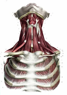
Background imageAnatomy Collection: Chest and neck muscles - 1866 illustration showing the structure of the muscles (brown)

Chest and neck muscles - 1866 illustration showing the structure of the muscles (brown) and tendons (white)
JAB4119416 Chest and neck muscles - 1866 illustration showing the structure of the muscles (brown) and tendons (white) found in the human neck (upper centre) and chest (bottom)

Background imageAnatomy Collection: Human skull - 1866 illustration showing the anatomy of the human skull - La head - Descriptive

Human skull - 1866 illustration showing the anatomy of the human skull - La head - Descriptive anatomy - Fig 1
JAB4119396 Human skull - 1866 illustration showing the anatomy of the human skull - La head - Descriptive anatomy - Fig 1: Region laterale, Fig 2: Region superieure

Background imageAnatomy Collection: Leg joints - 1866 anatomical illustration showing the tendons (white) and bones (beige)

Leg joints - 1866 anatomical illustration showing the tendons (white) and bones (beige) of the human hip (top)
JAB4119386 Leg joints - 1866 anatomical illustration showing the tendons (white) and bones (beige) of the human hip (top) and knee (bottom) joints - Pelvis bone joints - Fig 1

Background imageAnatomy Collection: Knee and ankle joints - 1866 anatomical illustration showing the tendons (white) and bones (beige)

Knee and ankle joints - 1866 anatomical illustration showing the tendons (white) and bones (beige)
JAB4119379 Knee and ankle joints - 1866 anatomical illustration showing the tendons (white) and bones (beige) that make up the human knee and ankle joints- Fig 1 and 2

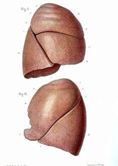
Background imageAnatomy Collection: Lung anatomy - 1866 illustration showing the external structure of the left (top) and right (bottom)

Lung anatomy - 1866 illustration showing the external structure of the left (top) and right (bottom)
JAB4119370 Lung anatomy - 1866 illustration showing the external structure of the left (top) and right (bottom) lungs - Lung - Descriptive anatomy - Fig 1: right lung, outer face, Fig 2: left lung

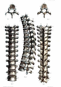
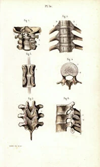
Background imageAnatomy Collection: Thoracic spine anatomy - 1866 anatomical illustration of the thoracic spine

Thoracic spine anatomy - 1866 anatomical illustration of the thoracic spine
JAB4119368 Thoracic spine anatomy - 1866 anatomical illustration of the thoracic spine, showing the individual vertebrae - Vertebral column - Dorsal vertebrae, osteology, locomotion

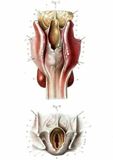
Background imageAnatomy Collection: Human throat 1866 cutaway illustration showing the anatomy of the throat LE LARYNX

Human throat 1866 cutaway illustration showing the anatomy of the throat LE LARYNX
JAB4119196 Human throat 1866 cutaway illustration showing the anatomy of the throat LE LARYNX, posterieure face - Fig 1: Pharynx and esophagus, Fig 2: Opening of the larynx 5 sup-glottic part

Background imageAnatomy Collection: Voice box anatomy 1866 anatomical illustration showing the muscles (brown)

Voice box anatomy 1866 anatomical illustration showing the muscles (brown) and external (Figs 1 and 2)
JAB4119181 Voice box anatomy 1866 anatomical illustration showing the muscles (brown) and external (Figs 1 and 2) and internal structure (Fig 3) of the larynx (voice box) Muscles du larynx - Fig 1

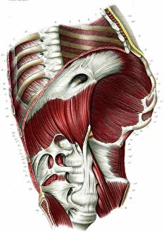
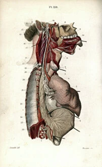
Background imageAnatomy Collection: Diaphragm 1866

Diaphragm 1866
JAB4119165 Diaphragm 1866; (add.info.: Diaphragm 1866 illustration of a section through the abdomen and chest, showing the diaphragm (arched)

Background imageAnatomy Collection: Menstrual cycle anatomy 1866

Menstrual cycle anatomy 1866
JAB4119155 Menstrual cycle anatomy 1866; (add.info.: Menstrual cycle anatomy 1866 illustrations showing the internal anatomy of a human ovary containing Graafian (ovarian) follicles (Fig 1)

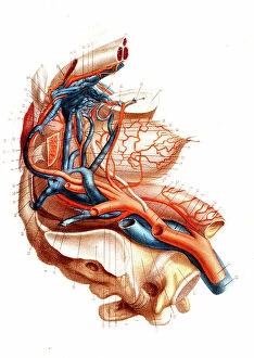
Background imageAnatomy Collection: Male pelvic blood supply 1866 illustration showing the major veins (blue) and arteries (red)

Male pelvic blood supply 1866 illustration showing the major veins (blue) and arteries (red)
JAB4119143 Male pelvic blood supply 1866 illustration showing the major veins (blue) and arteries (red) of the male pelvis These include the inferior vena cava, common iliac vein

Background imageAnatomy Collection: BARTHOLIN, Thomas (1616-1680). Danish physician, mathematician, and theologian

BARTHOLIN, Thomas (1616-1680). Danish physician, mathematician, and theologian
CUL4875648 BARTHOLIN, Thomas (1616-1680). Danish physician, mathematician, and theologian. He is best known for his work in the discovery of the lymphatic system in humans

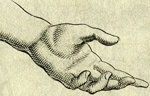
Background imageAnatomy Collection: study of a body, hand, copperplate engraving, about 1700

study of a body, hand, copperplate engraving, about 1700
UTD4921972 study of a body, hand, copperplate engraving, about 1700; (add.info.: study of a body, hand, copperplate engraving)

Background imageAnatomy Collection: The eye , copperplate engraving, about 1700

The eye , copperplate engraving, about 1700
UTD4921822 The eye , copperplate engraving, about 1700; (add.info.: The eye , copperplate engraving, about 1700 ©HistAuge/United Archives/Leemage); United Archives/HistAuge.

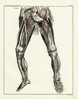
Background imageAnatomy Collection: Anatomy of the human arterial system in the legs. Handcoloured lithograph by St

Anatomy of the human arterial system in the legs. Handcoloured lithograph by St
FLO4684812 Anatomy of the human arterial system in the legs. Handcoloured lithograph by St. Schillinger from Lorenz Oken's Universal Natural History, Allgemeine Naturgeschichte fur alle Stande

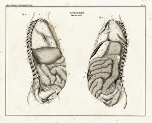
Background imageAnatomy Collection: Anatomy of human intestines from the side. Lithograph from Lorenz Oken's Universal Natural History

Anatomy of human intestines from the side. Lithograph from Lorenz Oken's Universal Natural History
FLO4684807 Anatomy of human intestines from the side. Lithograph from Lorenz Oken's Universal Natural History, Allgemeine Naturgeschichte fur alle Stande, Stuttgart

Background imageAnatomy Collection: Human skeleton from the front. Engraving by Fournier after an illustration by Martin St

Human skeleton from the front. Engraving by Fournier after an illustration by Martin St
FLO4685437 Human skeleton from the front. Engraving by Fournier after an illustration by Martin St. Ange from Charles d'Orbigny's Dictionary of Natural History, Paris
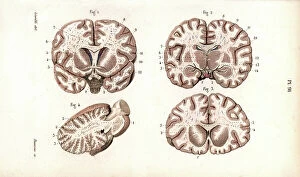
Background imageAnatomy Collection: Brain sections. Lithograph by Davesne, based on a drawing by Leveille

Brain sections. Lithograph by Davesne, based on a drawing by Leveille, in Petity Atlas complet d'Anatomie
FLO4673204 Brain sections. Lithograph by Davesne, based on a drawing by Leveille, in Petity Atlas complet d'Anatomie descriptive du Corps Humain, by Dr

Background imageAnatomy Collection: Vertebral column, ribs, vertebrae and sternum. Lithograph of Corbie

Vertebral column, ribs, vertebrae and sternum. Lithograph of Corbie, in Petity Atlas complet d'Anatomie descriptive
FLO4673029 Vertebral column, ribs, vertebrae and sternum. Lithograph of Corbie, in Petity Atlas complet d'Anatomie descriptive du Corps Humain, by Dr

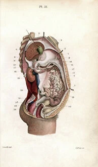
Background imageAnatomy Collection: Section through the abdomen. Lithograph of Lebrun, based on a drawing by Leveille

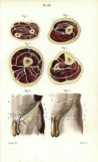
Section through the abdomen. Lithograph of Lebrun, based on a drawing by Leveille
FLO4672840 Section through the abdomen. Lithograph of Lebrun, based on a drawing by Leveille, in Petity Atlas complet d'Anatomie descriptive du Corps Humain, by Dr

Background imageAnatomy Collection: Aponevrosis (fibrous membrane that covers and supports the muscles) of the torso

Aponevrosis (fibrous membrane that covers and supports the muscles) of the torso, cross section and frontal view
FLO4672748 Aponevrosis (fibrous membrane that covers and supports the muscles) of the torso, cross section and frontal view

Background imageAnatomy Collection: Aponevrosis of the neck, shoulder and eye. Lithograph of Corbie, based on a drawing by Leveille

Aponevrosis of the neck, shoulder and eye. Lithograph of Corbie, based on a drawing by Leveille
FLO4672728 Aponevrosis of the neck, shoulder and eye. Lithograph of Corbie, based on a drawing by Leveille, in Petiure Atlas complet d'Anatomie descriptive du Corps Humain, by Dr

Background imageAnatomy Collection: Muscles and tendons of the leg and foot. Lithograph of Lebrun, based on a drawing by Leveille

Muscles and tendons of the leg and foot. Lithograph of Lebrun, based on a drawing by Leveille
FLO4672719 Muscles and tendons of the leg and foot. Lithograph of Lebrun, based on a drawing by Leveille, in Petity Atlas complet d'Anatomie descriptive du Corps Humain, by Dr

Background imageAnatomy Collection: Thigh and knee muscles and tendons. Lithograph of Lebrun, based on a drawing by Leveille

Thigh and knee muscles and tendons. Lithograph of Lebrun, based on a drawing by Leveille
FLO4672703 Thigh and knee muscles and tendons. Lithograph of Lebrun, based on a drawing by Leveille, in Petity Atlas complet d'Anatomie descriptive du Corps Humain, by Dr

Background imageAnatomy Collection: Thigh muscles and tendons. Lithograph of Lebrun, based on a drawing by Leveille

Thigh muscles and tendons. Lithograph of Lebrun, based on a drawing by Leveille
FLO4672702 Thigh muscles and tendons. Lithograph of Lebrun, based on a drawing by Leveille, in Petity Atlas complet d'Anatomie descriptive du Corps Humain, by Dr

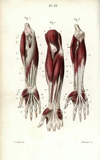
Background imageAnatomy Collection: Muscles and tendons of the forearm and wrist. Lithograph by Davesne, based on a drawing by Leveille

Muscles and tendons of the forearm and wrist. Lithograph by Davesne, based on a drawing by Leveille
FLO4672691 Muscles and tendons of the forearm and wrist. Lithograph by Davesne, based on a drawing by Leveille, in Petity Atlas complet d'Anatomie descriptive du Corps Humain, by Dr

Background imageAnatomy Collection: Muscles of the forearm and wrist. Lithograph by Davesne, based on a drawing by Leveille

Muscles of the forearm and wrist. Lithograph by Davesne, based on a drawing by Leveille
FLO4672685 Muscles of the forearm and wrist. Lithograph by Davesne, based on a drawing by Leveille, in Petity Atlas complet d'Anatomie descriptive du Corps Humain, by Dr

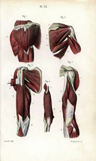
Background imageAnatomy Collection: Shoulder and arm muscles. Lithograph of Anneshower, based on a drawing by Leveille

Shoulder and arm muscles. Lithograph of Anneshower, based on a drawing by Leveille
FLO4672670 Shoulder and arm muscles. Lithograph of Anneshower, based on a drawing by Leveille, in Petiure Atlas complet d'Anatomie descriptive du Corps Humain, by Dr

Background imageAnatomy Collection: Internal muscles of the back. Lithograph by Davesne, based on a drawing by Leveille

Internal muscles of the back. Lithograph by Davesne, based on a drawing by Leveille
FLO4672652 Internal muscles of the back. Lithograph by Davesne, based on a drawing by Leveille, in Petity Atlas complet d'Anatomie descriptive du Corps Humain, by Dr

Background imageAnatomy Collection: Torso muscles (male). Lithograph of anneshower, based on a drawing by Leveille

Torso muscles (male). Lithograph of anneshower, based on a drawing by Leveille
FLO4672627 Torso muscles (male). Lithograph of anneshower, based on a drawing by Leveille, in Petiter Atlas complet d'Anatomie descriptive du Corps Humain, by Dr

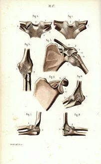
Background imageAnatomy Collection: Shoulder and elbow joints. Lithograph of Corbie, in Petity Atlas complet d'Anatomie descriptive du

Shoulder and elbow joints. Lithograph of Corbie, in Petity Atlas complet d'Anatomie descriptive du Corps Humain
FLO4672545 Shoulder and elbow joints. Lithograph of Corbie, in Petity Atlas complet d'Anatomie descriptive du Corps Humain, by Dr

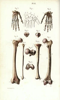
Background imageAnatomy Collection: Hand bone: carps, metacarps and phalanges and leg: femur and shin. Lithograph of Corbie

Hand bone: carps, metacarps and phalanges and leg: femur and shin. Lithograph of Corbie
FLO4672512 Hand bone: carps, metacarps and phalanges and leg: femur and shin. Lithograph of Corbie, in Petity Atlas complet d'Anatomie descriptive du Corps Humain, by Dr

Background imageAnatomy Collection: Cervical plexus. Lithograph of Corbie, based on a drawing by Leveille

Cervical plexus. Lithograph of Corbie, based on a drawing by Leveille, in Petity Atlas complet d'Anatomie
FLO4673165 Cervical plexus. Lithograph of Corbie, based on a drawing by Leveille, in Petity Atlas complet d'Anatomie descriptive du Corps Humain, by Dr

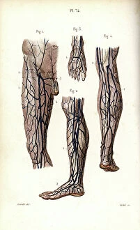
Background imageAnatomy Collection: Veins of the legs and foot. Lithograph of Corbie, based on a drawing by Leveille

Veins of the legs and foot. Lithograph of Corbie, based on a drawing by Leveille
FLO4673005 Veins of the legs and foot. Lithograph of Corbie, based on a drawing by Leveille, in Petity Atlas complet d'Anatomie descriptive du Corps Humain, by Dr

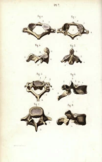
Background imageAnatomy Collection: Axis and vertebrae. Lithograph of Corbie, in Petity Atlas complet d'Anatomie descriptive du Corps

Axis and vertebrae. Lithograph of Corbie, in Petity Atlas complet d'Anatomie descriptive du Corps Humain, by Dr
FLO4672967 Axis and vertebrae. Lithograph of Corbie, in Petity Atlas complet d'Anatomie descriptive du Corps Humain, by Dr

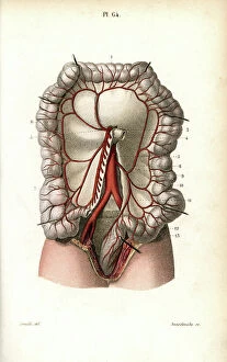
Background imageAnatomy Collection: Circulatory system of the intestines. Lithograph of Anneshower, based on a drawing by Leveille

Circulatory system of the intestines. Lithograph of Anneshower, based on a drawing by Leveille
FLO4672922 Circulatory system of the intestines. Lithograph of Anneshower, based on a drawing by Leveille, in Petity Atlas complet d'Anatomie descriptive du Corps Humain, by Dr

Background imageAnatomy Collection: Back and torso muscles. Lithograph by Davesne, based on a drawing by Leveille

Back and torso muscles. Lithograph by Davesne, based on a drawing by Leveille
FLO4672626 Back and torso muscles. Lithograph by Davesne, based on a drawing by Leveille, in Petity Atlas complet d'Anatomie descriptive du Corps Humain, by Dr

Background imageAnatomy Collection: Pelvic bone, front and back views. Lithograph of Corbie, in Petity Atlas complet d'Anatomie

Pelvic bone, front and back views. Lithograph of Corbie, in Petity Atlas complet d'Anatomie descriptive du Corps
FLO4672563 Pelvic bone, front and back views. Lithograph of Corbie, in Petity Atlas complet d'Anatomie descriptive du Corps Humain, by Dr

Background imageAnatomy Collection: Vertebral column, spine and neck. Lithograph of Corbie, in Petity Atlas complet d'Anatomie

Vertebral column, spine and neck. Lithograph of Corbie, in Petity Atlas complet d'Anatomie descriptive du Corps
FLO4672546 Vertebral column, spine and neck. Lithograph of Corbie, in Petity Atlas complet d'Anatomie descriptive du Corps Humain, by Dr

Background imageAnatomy Collection: Glossopharyngeal nerve. Lithograph by Davesne, based on a drawing by Leveille

Glossopharyngeal nerve. Lithograph by Davesne, based on a drawing by Leveille
FLO4672515 Glossopharyngeal nerve. Lithograph by Davesne, based on a drawing by Leveille, in Petity Atlas complet d'Anatomie descriptive du Corps Humain, by Dr

Background imageAnatomy Collection: Nerves of the skull and eye. Lithograph by Davesne, based on a drawing by Leveille

Nerves of the skull and eye. Lithograph by Davesne, based on a drawing by Leveille
FLO4672497 Nerves of the skull and eye. Lithograph by Davesne, based on a drawing by Leveille, in Petity Atlas complet d'Anatomie descriptive du Corps Humain, by Dr

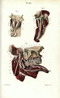
Background imageAnatomy Collection: Ophthalmic and maxillary nerves of the fifth cranial nerve. Lithograph by Davesne

Ophthalmic and maxillary nerves of the fifth cranial nerve. Lithograph by Davesne, based on a drawing by Leveille
FLO4672496 Ophthalmic and maxillary nerves of the fifth cranial nerve. Lithograph by Davesne, based on a drawing by Leveille, in Petity Atlas complet d'Anatomie descriptive du Corps Humain


All Professionally Made to Order for Quick Shipping

"Unveiling the Intricacies of Anatomy: From Sensory Homunculus to Motor Homunculus" Step into the fascinating world of anatomy, where every detail tells a story. Explore the depths of human and animal structures as we delve into this captivating subject. Let's begin with Leonardo da Vinci's masterpiece, "Head of a Bear. " Created in 1480 but discovered in 1945, this artwork showcases da Vinci's unparalleled ability to capture anatomical accuracy. The intricate details reveal his keen observation skills and deep understanding of form. Moving on to our furry friends, the greyhound takes center stage. Discovering the anatomy of these majestic creatures unveils their remarkable speed and agility. From their sleek skeleton to their muscular build, it is evident why they are renowned for their racing abilities. But what about us humans? Enter the sensory homunculus – a visual representation that depicts how our brain perceives different body parts based on sensitivity levels. This map reveals intriguing insights into how our senses are distributed throughout our bodies. X-ray images provide another dimension to understanding anatomy. Take a glimpse at normal knees through an X-ray lens – marvel at the complexity hidden beneath our skin while appreciating its delicate balance between strength and flexibility. The backbone is often considered one of nature's most ingenious designs. Our human backbone includes ribs and pelvis, providing stability while allowing movement – truly an architectural marvel worth exploring further. Venturing deeper within ourselves, let us explore the intricacies of the human brain from an inferior view. Witness its complex network of connections responsible for controlling various bodily functions - truly awe-inspiring. As we journey back in time, we encounter hominid crania – remnants that shed light on our evolutionary history. These ancient skulls offer glimpses into early forms such as Australopithecus afarensis (AL 288-1), famously known as Lucy - bridging gaps between past and present.