Fraud Blocker Skip to main content

Angiogram Collection

"Exploring the Intricate Network of Blood Vessels

Background imageAngiogram Collection: Brain blood vessels, 3D angiogram C007 / 1981

Brain blood vessels, 3D angiogram C007 / 1981
Brain blood vessels. Coloured 3D lateral angiogram (blood vessel X-ray) of the blood vessels in the brain of a healthy 32-year-old

Background imageAngiogram Collection: Coronary arteriogram of arteries of the heart 1904

Coronary arteriogram of arteries of the heart 1904
Black-And-White, Black and White, Monochrome Image, Diagnosis, X-Ray, Historical, X-Ray Photography, Early 1904, Coronary Angiography, 1904, Early, Heart Arteries, Arteriography, Coronary 1904

Background imageAngiogram Collection: Ischaemia, digital angiogram

Ischaemia, digital angiogram
image d angiographie numerisee( non soustraite) de la main gauche, en vue de face, par catheterisme hyperselectif des arteres du membre superieur gauche, sous anasthesie generale

Background imageAngiogram Collection: Brain aneurysm, 3D scan

Brain aneurysm, 3D scan
Brain aneurysm. 3D computed tomography (CT) angiogram coupled with a magnetic resonance imaging (MRI) scan of the brain of a 38-year-old, showing a large aneurysm (bright)

Background imageAngiogram Collection: Vascular system of the head, MRI scan

Vascular system of the head, MRI scan
Vascular system of the head. Magnetic resonance imaging (MRI) angiogram of a sagittal section through the head of a healthy 43-year-old, showing the blood vessels (white) of the brain and neck

Background imageAngiogram Collection: Neck and head arteries, MRA scan C016 / 6449

Neck and head arteries, MRA scan C016 / 6449
Neck and head arteries. Coloured magnetic resonance angiography (MRA) scan of a normal carotid system of a 36 year old patient

Background imageAngiogram Collection: Balloon angioplasty, X-ray

Balloon angioplasty, X-ray
Balloon angioplasty. Coloured X-ray of the abdomen of a 45-year-old patient with a history of thrombosis (blood clots) in the inferior vena cava (vein), which have narrowed (stenosed)

Background imageAngiogram Collection: External carotid artery, 3D CT scan C016 / 6341

External carotid artery, 3D CT scan C016 / 6341
External carotid artery. Coloured 3D computed tomography (CT) angiogram (blood vessel scan) of the head showing a normal external carotid artery (red, in neck) and its branches

Background imageAngiogram Collection: Artery damage, MRA scan

Artery damage, MRA scan
Artery damage. Magnetic resonance angiography (MRA) scan of the neck of a 23-year-old male motorcycle crash victim. Dissection of the right internal carotid artery (left)

Background imageAngiogram Collection: Coronary stent, X-ray F006 / 9141

Coronary stent, X-ray F006 / 9141
Coronary stent. Coloured angiogram (blood vessel X-ray) of the coronary arteries of a 52 year old patient. The artery at right has had a stent placed in it to treat a blockage

Background imageAngiogram Collection: Aorta, 3D CT scan F006 / 9120

Aorta, 3D CT scan F006 / 9120
Aorta. Coloured 3D computed tomography (CT) angiogram of the aorta of a 54 year old patient. The aorta is the bodys main artery

Background imageAngiogram Collection: Aorta, 3D CT scan F006 / 9119

Aorta, 3D CT scan F006 / 9119
Aorta. Coloured 3D computed tomography (CT) angiogram of the aorta of a 54 year old patient. The aorta is the bodys main artery

Background imageAngiogram Collection: Narrowed arteries, 3D CT scan F006 / 9113

Narrowed arteries, 3D CT scan F006 / 9113
Narrowed arteries. Coloured 3D computer tomography (CT) angiogram of the pelvis of a 66 year old male patient with narrowing (stenosis) of the iliac and femoral arteries

Background imageAngiogram Collection: Abdominal aorta, 3D CT scan F006 / 9102

Abdominal aorta, 3D CT scan F006 / 9102
Abdominal aorta. Coloured 3D computed tomography (CT) angiogram of the abdominal aorta of a 53 year old patient. The aorta is the bodys main artery

Background imageAngiogram Collection: Abdominal aorta, 3D CT scan F006 / 9101

Abdominal aorta, 3D CT scan F006 / 9101
Abdominal aorta. Coloured 3D computed tomography (CT) angiogram of the abdominal aorta of a 53 year old patient. The aorta is the bodys main artery

Background imageAngiogram Collection: Abdominal aorta, 3D CT scan F006 / 9095

Abdominal aorta, 3D CT scan F006 / 9095
Abdominal aorta. Coloured 3D computed tomography (CT) angiogram of the pelvis of a 39 year old patient. At top is the abdominal aorta. The aorta is the bodys main artery

Background imageAngiogram Collection: Abdominal aorta, 3D CT scan F006 / 9094

Abdominal aorta, 3D CT scan F006 / 9094
Abdominal aorta, 3D CT scan

Background imageAngiogram Collection: Abdominal aorta, 3D CT scan F006 / 9091

Abdominal aorta, 3D CT scan F006 / 9091
Abdominal aorta. Coloured 3D computed tomography (CT) angiogram of the abdominal aorta in a 42 year old patient. The aorta is the bodys main artery

Background imageAngiogram Collection: Abdominal aorta, 3D CT scan F006 / 9089

Abdominal aorta, 3D CT scan F006 / 9089
Abdominal aorta. Coloured 3D computed tomography (CT) angiogram of the abdominal aorta in a 42 year old patient. The aorta is the bodys main artery

Background imageAngiogram Collection: Thrombophlebitis in the brain, MRI scans

Thrombophlebitis in the brain, MRI scans
Thrombophlebitis in the brain. Coloured magnetic resonance imaging (MRI) angiograms comparing the blood vessels (white) of the brain and neck in a healthy 43-year-old (left)

Background imageAngiogram Collection: Brain aneurysm, DSA scan

Brain aneurysm, DSA scan
Brain aneurysm. Coloured digital subtraction angiogram (DSA) of the head of a 38-year-old, showing a large aneurysm (bright, centre) of the right internal carotid artery

Background imageAngiogram Collection: Thrombophlebitis in the brain, 3D CT scan

Thrombophlebitis in the brain, 3D CT scan
Thrombophlebitis in the brain. Coloured 3D computed tomography (CT) angiogram of the blood vessels (blue) in the brain of a 48-year-old with thrombophlebitis of the superior sagittal sinus

Background imageAngiogram Collection: Atherosclerosis, 3D CT scan

Atherosclerosis, 3D CT scan
Atherosclerosis. 3D computed tomography (CT) angiogram of the main blood vessels of the lower body a 58-year-old man, showing numerous atheromatous plaques (light, centre) on the vessel walls

Background imageAngiogram Collection: Atherosclerosis, 3D CT scan C016 / 6537

Atherosclerosis, 3D CT scan C016 / 6537
Atherosclerosis. Coloured 3D computed tomography (CT) angiogram of the hip of a 67 year old patient with hypertension (high blood pressure) and hypercholesterolemia (high blood cholesterol)

Background imageAngiogram Collection: Atherosclerosis, 3D CT scan C016 / 6540

Atherosclerosis, 3D CT scan C016 / 6540
Atherosclerosis. Coloured 3D computed tomography (CT) angiogram of the abdomen of a 73 year old patient. The aorta (cream, down centre), the bodys main artery, has calcified fatty plaques (atheroma)

Background imageAngiogram Collection: Atherosclerosis, 3D CT scan C016 / 6499

Atherosclerosis, 3D CT scan C016 / 6499
Atherosclerosis. Coloured 3D computed tomography (CT) angiogram of the head of a 60 year old patient with hypertension (high blood pressure) and hypercholesterolemia (high blood cholesterol)

Background imageAngiogram Collection: Neck arthritis treatment, angiogram C016 / 6440

Neck arthritis treatment, angiogram C016 / 6440
Neck arthritis treatment. Coloured X-ray angiogram of the neck of a 48 year old patient with severe osteoarthritis of the neck

Background imageAngiogram Collection: Neck arthritis treatment, angiogram C016 / 6443

Neck arthritis treatment, angiogram C016 / 6443
Neck arthritis treatment. Coloured X-ray angiogram of the neck of a 48 year old patient with severe osteoarthritis of the neck

Background imageAngiogram Collection: Atherosclerosis, 3D CT scan C016 / 6538

Atherosclerosis, 3D CT scan C016 / 6538
Atherosclerosis. Coloured 3D computed tomography (CT) angiogram of the hip of a 67 year old patient with hypertension (high blood pressure) and hypercholesterolemia (high blood cholesterol)

Background imageAngiogram Collection: Atherosclerosis, 3D CT scan C016 / 6539

Atherosclerosis, 3D CT scan C016 / 6539
Atherosclerosis. Coloured 3D computed tomography (CT) angiogram of the heart of a 69 year old patient that has had an aortic heart valve replacement

Background imageAngiogram Collection: Portal vein surgery, X-ray C016 / 6546

Portal vein surgery, X-ray C016 / 6546
Vein surgery. Coloured angiogram (X-ray) of surgery to lower the blood pressure in the portal vein of a 60 year old patient

Background imageAngiogram Collection: Internal carotid artery, 3D CT scan C016 / 6399

Internal carotid artery, 3D CT scan C016 / 6399
Internal carotid artery. Coloured 3D computed tomography (CT) angiogram (blood vessel scan) of the head showing a normal internal carotid artery (red, in neck) and its intra-cerebral branches

Background imageAngiogram Collection: Portal vein surgery, X-ray C016 / 6543

Portal vein surgery, X-ray C016 / 6543
Vein surgery. Angiogram (X-ray) of surgery to lower the blood pressure in the portal vein of a 60 year old patient. A catheter (white line running down from top centre)

Background imageAngiogram Collection: Internal carotid artery, 3D CT scan C016 / 6400

Internal carotid artery, 3D CT scan C016 / 6400
Internal carotid artery. Coloured 3D computed tomography (CT) angiogram (blood vessel scan) of the head showing a normal internal carotid artery (red, in neck) and its intra-cerebral branches

Background imageAngiogram Collection: Portal vein surgery, X-ray C016 / 6545

Portal vein surgery, X-ray C016 / 6545
Vein surgery. Coloured angiogram (X-ray) of surgery to lower the blood pressure in the portal vein of a 60 year old patient

Background imageAngiogram Collection: Portal vein surgery, X-ray C016 / 6544

Portal vein surgery, X-ray C016 / 6544
Vein surgery. Coloured angiogram (X-ray) of surgery to lower the blood pressure in the portal vein of a 60 year old patient

Background imageAngiogram Collection: Blood supply of the hand, angiograms C018 / 0389

Blood supply of the hand, angiograms C018 / 0389
Blood supply of the hand. Coloured angiograms (X-rays) of the blood vessels of the right hand of a 65 year old patient with Dupuytrens contracture

Background imageAngiogram Collection: Forearm veins, X-ray C018 / 0475

Forearm veins, X-ray C018 / 0475
Forearm veins. Coloured venogram (vein X-ray) of the veins in the forearms of a 43 year old patient in need of dialysis. This venogram has been taken to evaluate the health of the veins prior to

Background imageAngiogram Collection: Aneurysm treatment, angiogram C018 / 0537

Aneurysm treatment, angiogram C018 / 0537
Aneurysm treatment. Coloured angiogram (X-ray) overlaid on a computer model of the treatment of a 39 year old patient with an aneurysm of the posterior communicating artery

Background imageAngiogram Collection: Aneurysm treatment, angiogram C018 / 0536

Aneurysm treatment, angiogram C018 / 0536
Aneurysm treatment. Angiogram (X-ray) overlaid on a computer model of the treatment of a 39 year old patient with an aneurysm of the posterior communicating artery

Background imageAngiogram Collection: Blood supply of the hand, angiogram C018 / 0387

Blood supply of the hand, angiogram C018 / 0387
Blood supply of the hand. Coloured angiogram (X-ray) of the blood vessels of the right hand of a 65 year old patient with Dupuytrens contracture

Background imageAngiogram Collection: Thrombophlebitis, angiogram C018 / 0529

Thrombophlebitis, angiogram C018 / 0529
Thrombophlebitis. Coloured angiogram (X-ray) of the brain of a 28 year old female patient who had thrombophelibitis of the superior sagittal sinus (indicated by arrows)

Background imageAngiogram Collection: Pinned broken leg, angiogram C018 / 0392

Pinned broken leg, angiogram C018 / 0392
Pinned broken leg. Coloured angiogram of the right leg of a 22 year old patient that sustained an open tibial (shin bone) fracture in a motorcycle accident. The fracture has been aligned and pinned

Background imageAngiogram Collection: Aneurysm treatment, angiogram C018 / 0525

Aneurysm treatment, angiogram C018 / 0525
Aneurysm treatment. Coloured angiogram (X-ray) overlaid on a computer model of the treatment of a patient with an aneurysm of the internal carotid artery

Background imageAngiogram Collection: Pinned broken leg, angiogram C018 / 0391

Pinned broken leg, angiogram C018 / 0391
Pinned broken leg. Coloured angiogram of the right leg of a 22 year old patient that sustained an open tibial (shin bone) fracture in a motorcycle accident. The fracture has been aligned and pinned

Background imageAngiogram Collection: Aneurysm treatment, angiogram C018 / 0514

Aneurysm treatment, angiogram C018 / 0514
Aneurysm treatment. Coloured angiogram (X-ray) overlaid on a computer model of the treatment of a 39 year old patient with an aneurysm of the posterior communicating artery

Background imageAngiogram Collection: Thrombophlebitis, angiogram C018 / 0530

Thrombophlebitis, angiogram C018 / 0530
Thrombophlebitis. Coloured angiogram (X-ray) of the brain of a 28 year old female patient who had thrombophelibitis of the superior sagittal sinus (top left)

Background imageAngiogram Collection: Blood supply of the hand, angiogram C018 / 0386

Blood supply of the hand, angiogram C018 / 0386
Blood supply of the hand. Coloured angiogram (X-ray) of the blood vessels of the right hand of a 65 year old patient with Dupuytrens contracture


All Professionally Made to Order for Quick Shipping

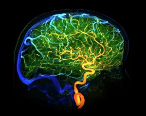
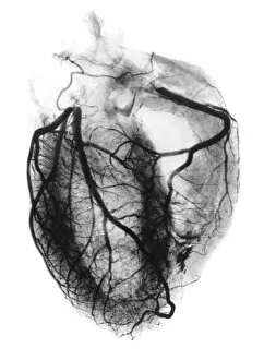
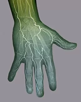
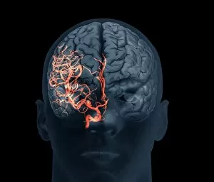
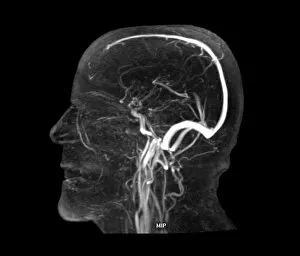
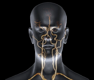
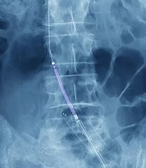
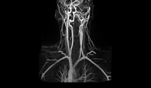
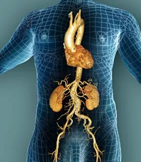
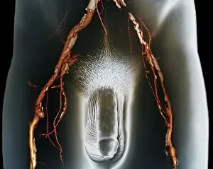
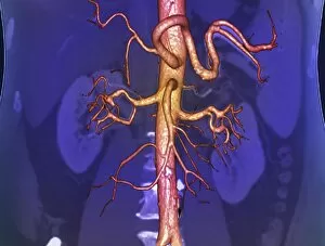
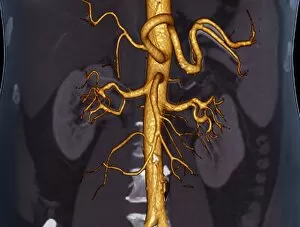
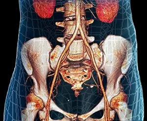
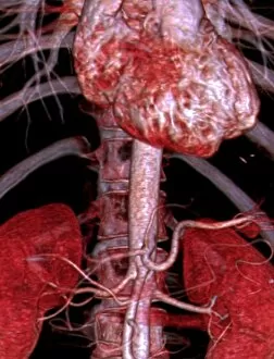
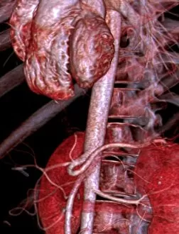
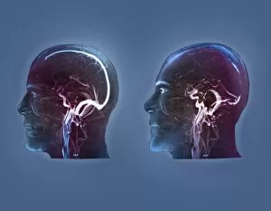
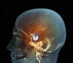
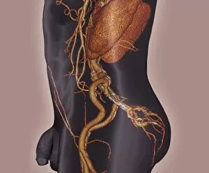
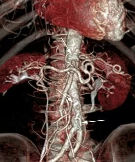
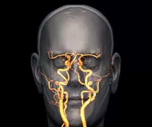
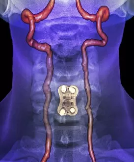
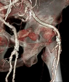
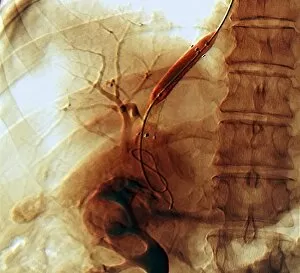
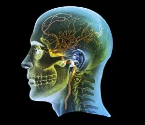
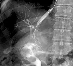
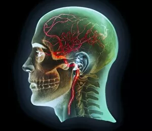
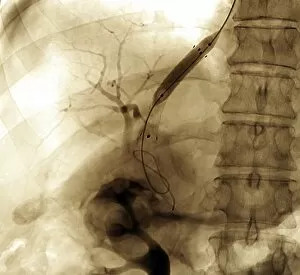
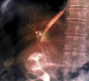
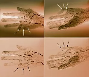
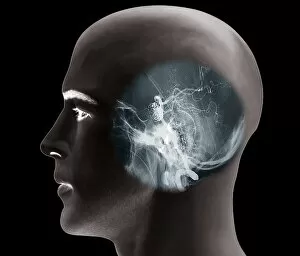
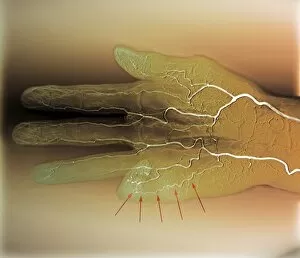
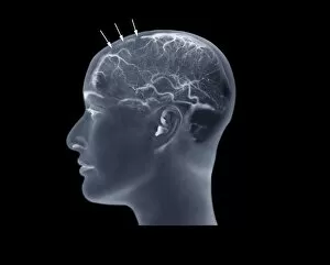
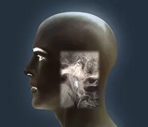
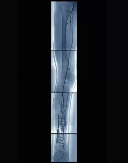
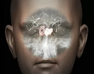
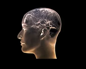
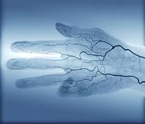
"Exploring the Intricate Network of Blood Vessels: Unveiling the Wonders of Angiogram" From unraveling the mysteries of brain blood vessels with a 3D angiogram C007 / 1981 to examining neck and shoulder arteries through X-ray, angiograms have revolutionized medical imaging. These powerful techniques allow us to visualize intricate pathways within our bodies, providing valuable insights into various conditions. With X-ray technology, we can delve into abdominal arteries using an X-ray P206 / 0309, enabling doctors to identify potential issues in this vital area. Moreover, body imaging allows for comprehensive assessments that aid in diagnosing complex ailments. Angiograms also play a crucial role in detecting cerebral vasculitis and assessing its severity through X-rays. Similarly, coronary arteriograms provide detailed images of heart arteries dating back as early as 1904, helping cardiologists diagnose and treat cardiovascular diseases effectively. Digital angiograms shed light on ischaemia by capturing precise images that highlight areas affected by reduced blood flow. Furthermore, cutting-edge technology enables us to examine brain aneurysms with remarkable clarity through advanced 3D scans. The vascular system of the head becomes visible like never before thanks to MRI scans specifically tailored for this purpose. This non-invasive technique aids in identifying any abnormalities or irregularities present. In cases requiring intervention, balloon angioplasty guided by X-rays proves invaluable. By visualizing the procedure's progress in real-time, physicians can restore proper blood flow efficiently. For a more detailed examination of external carotid artery anatomy and functionality, a three-dimensional CT scan C016 / 6341 offers unparalleled precision and accuracy. Lastly, MRA scans help detect artery damage caused by trauma or disease processes accurately. By utilizing magnetic resonance imaging techniques specialized for analyzing blood vessels' condition without invasive procedures such as surgery or catheterization. As medical advancements continue to push boundaries forward, angiograms remain a cornerstone in diagnosing and treating various vascular conditions.