
Angiography Collection
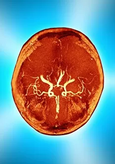
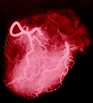
Angiography, a medical imaging technique, allows for the visualization of blood vessels in various parts of the body


All Professionally Made to Order for Quick Shipping
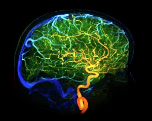
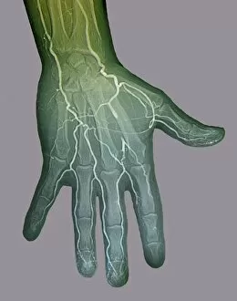
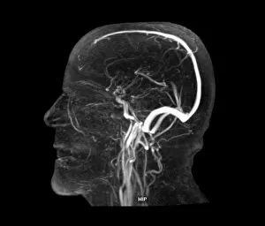
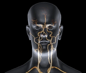
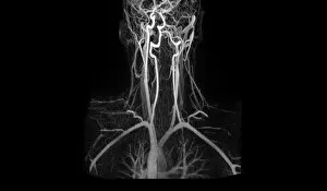
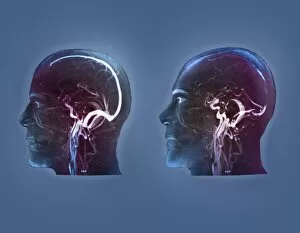
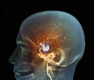
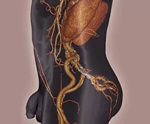
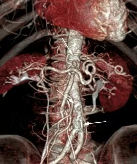
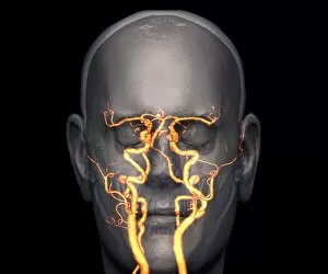
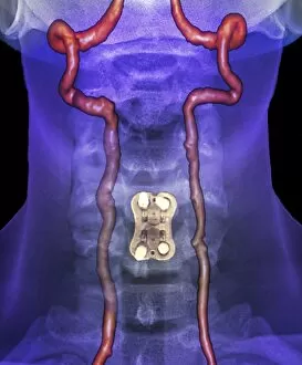
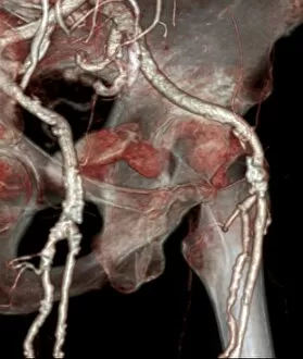
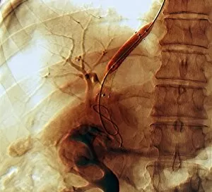
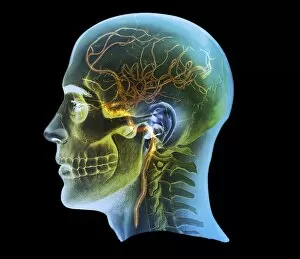
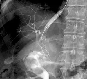
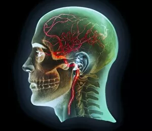
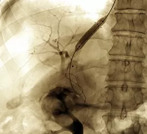
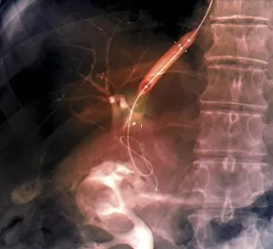
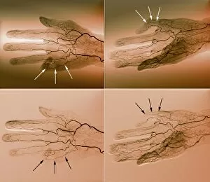
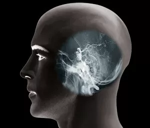
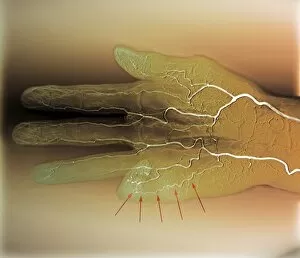
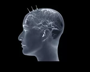
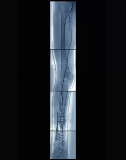
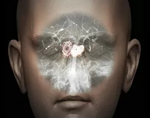
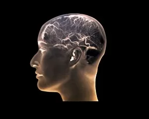
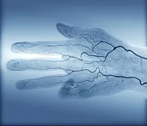
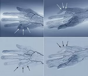
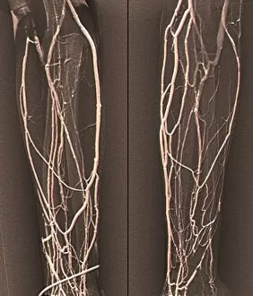
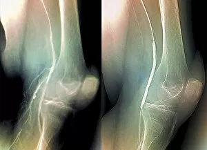
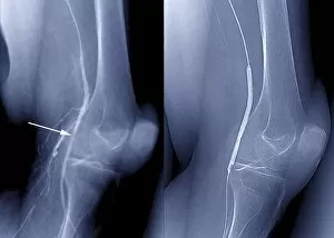
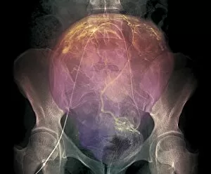
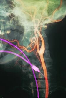
Angiography, a medical imaging technique, allows for the visualization of blood vessels in various parts of the body. From brain blood vessels to neck and shoulder arteries, this procedure provides valuable insights into our vascular system. In 1981, the advent of 3D angiogram C007 revolutionized our understanding of brain blood vessels by offering detailed images that were previously unimaginable. Moving down to the neck and shoulder region, X-ray angiography became instrumental in examining these crucial arteries. Similarly, abdominal arteries were explored using X-ray P206 / 0309 scans. These non-invasive techniques have proven invaluable in diagnosing conditions related to these vital areas. Beyond specific regions, it has expanded its reach to encompass body imaging as a whole. By utilizing digital angiograms and MRI scans like cerebral vasculitis or ischaemia detection methods, doctors can identify potential issues within the entire vascular system. Brain aneurysms are particularly well-suited for examination through angiographic techniques such as 3D scans or MRI scans focusing on the vascular system of the head. These advanced imaging technologies offer unprecedented clarity when it comes to detecting aneurysms and determining appropriate treatment plans. The external carotid artery's intricate structure was revealed through a groundbreaking 3D CT scan C016 / 6341. This innovation allowed medical professionals to gain comprehensive knowledge about this critical pathway supplying oxygenated blood to different regions of the head. Moreover, MRA scans have been pivotal in identifying artery damage caused by various factors such as trauma or disease progression. With their high-resolution capabilities, MRA scans enable physicians to make accurate diagnoses and develop effective treatment strategies accordingly. Lastly, thrombophlebitis within the brain has become more manageable due to advancements in MRI scanning technology specifically tailored for this condition. By capturing detailed images with exceptional precision during these specialized MRI scans, healthcare providers can promptly diagnose thrombophlebitis and initiate appropriate interventions.