Fraud Blocker Skip to main content

Antigenic Collection

"Unlocking the Power of Antigenic Interactions: Exploring T Lymphocytes and Cancer Cells" In the realm of immunology

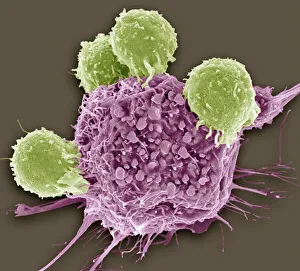
Background imageAntigenic Collection: T lymphocytes and cancer cell, SEM C001 / 1679

T lymphocytes and cancer cell, SEM C001 / 1679
T lymphocytes and cancer cell. Coloured scanning electron micrograph (SEM) of T lymphocyte cells (green) attached to a cancer cell

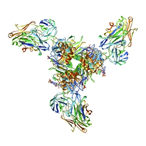
Background imageAntigenic Collection: Haemagglutinin viral surface protein F007 / 9932

Haemagglutinin viral surface protein F007 / 9932
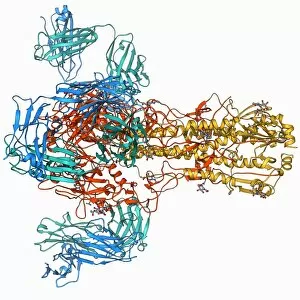
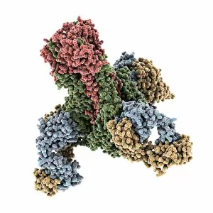
Haemagglutinin viral surface protein. Molecular model of haemagglutinin, a surface protein from the influenza virus, complexed with a neutralising antibody

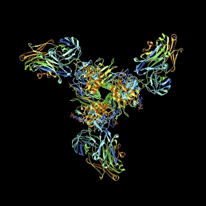
Background imageAntigenic Collection: Haemagglutinin viral surface protein F007 / 9931

Haemagglutinin viral surface protein F007 / 9931
Haemagglutinin viral surface protein. Molecular model of haemagglutinin, a surface protein from the influenza virus, complexed with a neutralising antibody

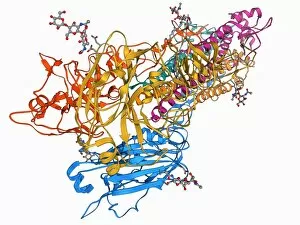
Background imageAntigenic Collection: H5N1 Haemagglutinin protein subunit F006 / 9590

H5N1 Haemagglutinin protein subunit F006 / 9590
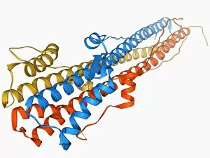
H5N1 haemagglutinin protein subunit. Molecular model of the haemagglutinin HA(5) subunit. Haemagglutinin is a surface protein from the influenza A virus

Background imageAntigenic Collection: Haemagglutinin protein subunit F006 / 9479

Haemagglutinin protein subunit F006 / 9479
Haemagglutinin protein subunit. Molecular model of the ectodomain of the haemagglutinin HA(2) subunit. Haemagglutinin is a surface protein from the influenza A virus

Background imageAntigenic Collection: Haemagglutinin viral surface protein F006 / 9470

Haemagglutinin viral surface protein F006 / 9470
Haemagglutinin viral surface protein. Molecular model of haemagglutinin, a surface protein from the influenza virus, complexed with a neutralising antibody

Background imageAntigenic Collection: Haemagglutinin viral surface protein C015 / 9965

Haemagglutinin viral surface protein C015 / 9965
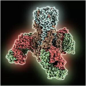
Haemagglutinin viral surface protein. Molecular model of haemagglutinin, a surface protein from the influenza virus, complexed with a neutralising antibody

Background imageAntigenic Collection: Haemagglutinin viral surface protein C015 / 7124

Haemagglutinin viral surface protein C015 / 7124
Haemagglutinin viral surface protein. Molecular model of haemagglutinin, a surface protein from the influenza virus, complexed with a neutralising antibody

Background imageAntigenic Collection: Haemagglutinin viral surface protein C015 / 9974

Haemagglutinin viral surface protein C015 / 9974
Haemagglutinin viral surface protein. Molecular model of haemagglutinin, a surface protein from the influenza virus, complexed with a neutralising antibody

Background imageAntigenic Collection: Haemagglutinin viral surface protein C015 / 7123

Haemagglutinin viral surface protein C015 / 7123
Haemagglutinin viral surface protein. Molecular model of haemagglutinin, a surface protein from the influenza virus, complexed with a neutralising antibody

Background imageAntigenic Collection: T lymphocytes and cancer cells, SEM

T lymphocytes and cancer cells, SEM
T lymphocytes and cancer cells. Coloured scanning electron micrograph (SEM) of T lymphocyte cells (purple) attached to cancer cells

Background imageAntigenic Collection: Influenza virus protein spikes

Influenza virus protein spikes. Computer artwork of part of the surface of a virus particle of the influenza (flu) virus. These spikes (formed by the surface proteins)

Background imageAntigenic Collection: Cd28 antigen molecule

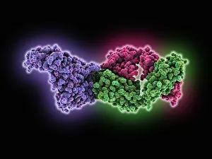
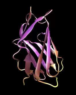
Cd28 antigen molecule. Computer model showing the secondary structure of cd28. Cd28 is an antigen found on the surface of T cells

Background imageAntigenic Collection: Influenza B virus neuraminidase enzyme

Influenza B virus neuraminidase enzyme
Neuraminidase enzyme, molecular model. This enzyme is found on the surface of the influenza B virus. It plays a role in releasing the viruses from infected cells once they have used the cells

Background imageAntigenic Collection: Haemagglutinin viral surface protein

Haemagglutinin viral surface protein, molecular model. Haemagglutinin is found on the surface of many bacteria and viruses, notably the influenza virus


All Professionally Made to Order for Quick Shipping

"Unlocking the Power of Antigenic Interactions: Exploring T Lymphocytes and Cancer Cells" In the realm of immunology, antigenic interactions play a pivotal role in understanding diseases like cancer. Recent studies have shed light on the fascinating relationship between T lymphocytes and cancer cells, revealing potential therapeutic avenues. SEM C001 / 1679 reveals intricate details of this interaction at a microscopic level, providing valuable insights into the immune response against malignant cells. The Haemagglutinin viral surface proteins F007 / 9932 and F007 / 9931 also contribute to our understanding by highlighting specific protein markers involved in these interactions. Delving deeper into influenza viruses, H5N1 Haemagglutinin protein subunit F006 / 9590 and its counterparts (F006 / 9479, F006 / 9470) hold significant importance. These viral surface proteins act as antigens that trigger an immune response within our bodies. The Haemagglutinin viral surface protein C015 family (C015/9965, C015/7124, C015/9974, C015/7123) further expands our knowledge base. Their unique structures aid in identifying virus strains while studying their antigenicity. Combining SEM images with research on T lymphocytes' behavior when confronting cancer cells provides invaluable information for developing targeted therapies. Understanding how these two entities interact can potentially unlock novel treatment strategies against various types of cancers. Moreover, exploring Influenza virus protein spikes adds another layer to this captivating field. By investigating their antigenic properties and mechanisms behind host-cell recognition, scientists strive to develop more effective vaccines capable of combating ever-evolving flu strains. The study interactions involving T lymphocytes and cancer cells offers hope for improved diagnostics and treatments.