
Bacteria Collection
"Bacteria: The Microscopic Marvels of Yellowstone National Park" In the heart of Wyoming, USA lies the breathtaking Yellowstone National Park


All Professionally Made to Order for Quick Shipping
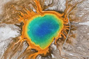
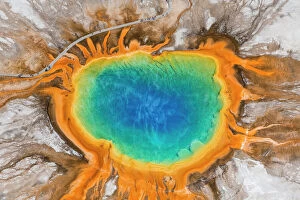
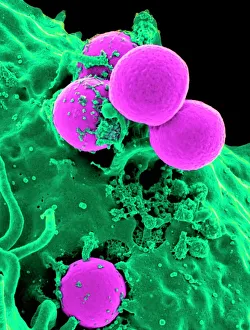
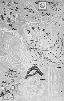
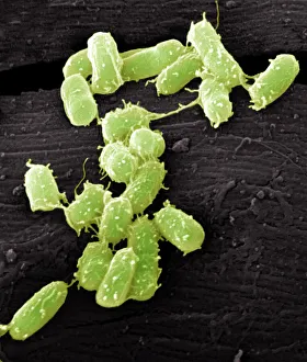
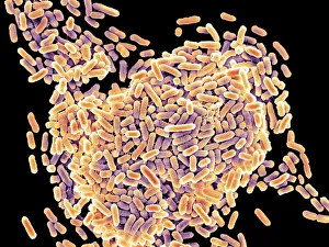
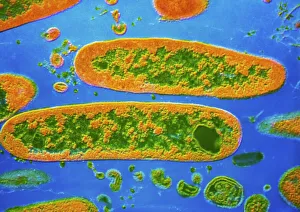
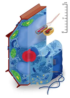
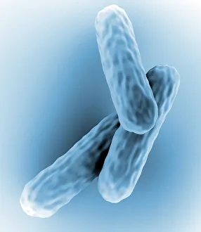
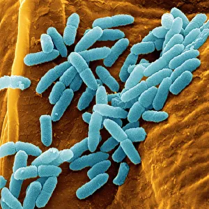
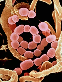
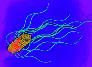
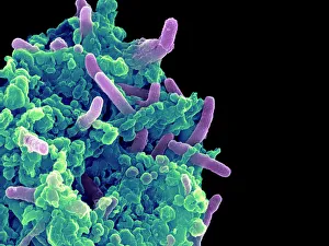
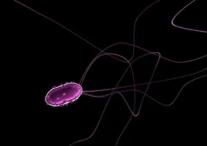
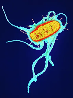
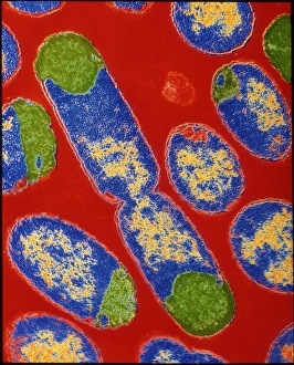
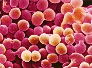
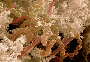
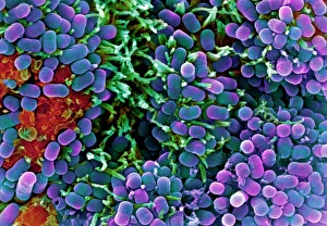
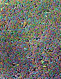
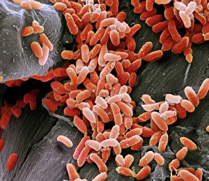
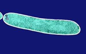
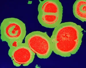
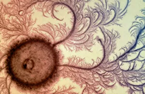
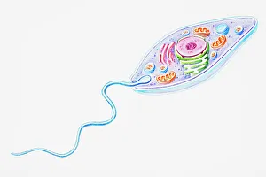
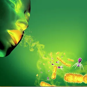
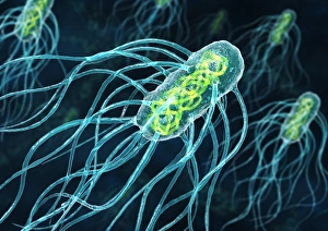
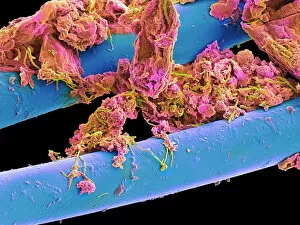
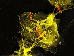
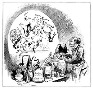
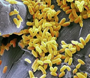
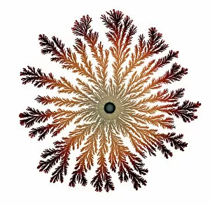
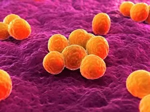
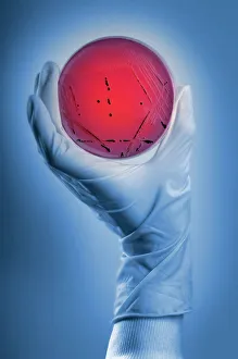
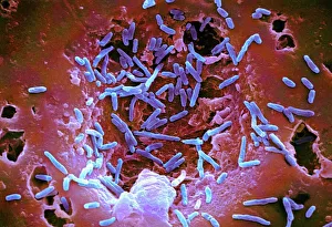
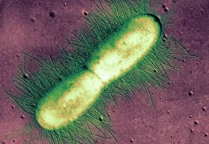
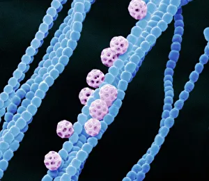
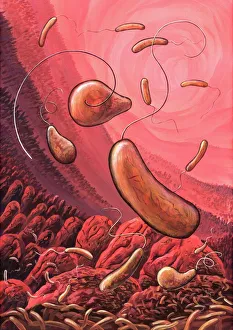
"Bacteria: The Microscopic Marvels of Yellowstone National Park" In the heart of Wyoming, USA lies the breathtaking Yellowstone National Park, a haven for nature enthusiasts and scientists alike. Amidst its natural wonders, one can find an extraordinary display of microbial life at the Grand Prismatic Spring in the Midway Geyser Basin. Picture No. 11675590 captures this mesmerizing sight, showcasing vibrant hues that seem to dance upon the water's surface. However, what may appear as an artistic masterpiece is actually a thriving community of bacteria. Zooming in closer with SEM C018 / 8596 reveals a neutrophil engulfing MRSA - Methicillin-resistant Staphylococcus aureus. This image serves as a reminder of how these tiny organisms can pose significant threats to human health. Another SEM image showcases E. Coli bacteria, reminding us that not all it can harmful; some play crucial roles in our digestive system and even aid scientific research. Moving back to Yellowstone's enchantment, we encounter Salmonella bacteria through another captivating SEM photograph. These microscopic creatures serve as a stark reminder that even within such serene landscapes, dangers lurk unseen. Coloured TEM imagery introduces us to Yersinia pestis bacteria – responsible for causing devastating outbreaks like the infamous Black Death throughout history. Such visuals emphasize their intricate structures and highlight why understanding them is vital for public health efforts. Venturing beyond real-life images into computer artwork brings forth a beta DNA segment surrounded by spheres—a representation of genetic material essential for bacterial survival and reproduction. Artistic depictions also shed light on various cell types found within these microorganisms' complex world—each playing unique roles necessary for their survival and proliferation. Historical diagrams reveal cultures of Anthrax—an infectious disease with severe consequences if left unchecked—underscoring humanity's ongoing battle against bacterial infections throughout time. Tuberculosis bacteria make their appearance too—a haunting reminder that despite medical advancements, infectious diseases continue to challenge us.