Fraud Blocker Skip to main content

Bacterial Collection (page 6)

"Bacterial: Unveiling the Microscopic World of Disease and Discovery" Step into a time capsule as we journey through history, exploring the captivating realm of bacteria

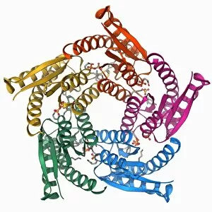
Background imageBacterial Collection: Lumazine synthase molecule F006 / 9291

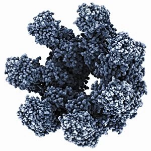
Lumazine synthase molecule F006 / 9291
Lumazine synthase molecule. Molecular model showing the structure of a lumazine synthase enzyme molecule from a Brucella abortus bacterium

Background imageBacterial Collection: Gene activator protein F006 / 9269

Gene activator protein F006 / 9269
Gene activator protein. Molecular model of catabolite gene activator protein (CAP, pink and green) bound to a molecule of deoxyribonucleic acid (DNA, across top)

Background imageBacterial Collection: E coli Holliday junction complex F006 / 9261

E coli Holliday junction complex F006 / 9261
E. coli Holliday junction complex. Molecular model of a RuvA protein (red) in complex with a Holliday junction between homologous strands of DNA (deoxyribonucleic acid, blue) from an E

Background imageBacterial Collection: Bioluminescent enzyme molecule F006 / 9256

Bioluminescent enzyme molecule F006 / 9256
Bioluminescent enzyme. Molecular model of the bioluminescent enzyme luciferase from the marine bacteria Vibrio harveyi

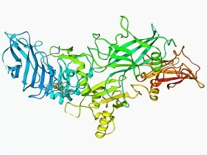
Background imageBacterial Collection: Anthrax protective antigen molecule F006 / 9229

Anthrax protective antigen molecule F006 / 9229
Anthrax protective antigen molecule. Computer model showing the structure of a molecule of protective antigen (PA) produced by anthrax (Bacillus anthracis) bacteria

Background imageBacterial Collection: Anthrax protective antigen molecule F006 / 9225

Anthrax protective antigen molecule F006 / 9225
Anthrax protective antigen molecule. Computer model showing the structure of a molecule of protective antigen (PA) produced by anthrax (Bacillus anthracis) bacteria

Background imageBacterial Collection: T-cell receptor bound to enterotoxin

T-cell receptor bound to enterotoxin, molecular model. The T cell receptor (TCR) is a protein complex found on the surface of a type of white blood cell called T lymphocytes (or T cells)

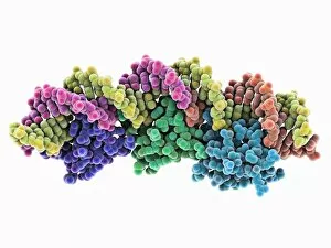
Background imageBacterial Collection: Excisionase complex with DNA

Excisionase complex with DNA. Molecular model of three excisionase proteins (bottom, purple, green and blue) bound to a strand of DNA (top, deoxyribonucleic acid)

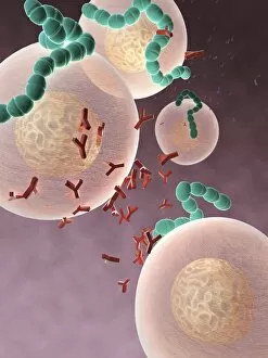
Background imageBacterial Collection: Antibodies and bacteria, artwork

Antibodies and bacteria, artwork
Antibodies and bacteria. Computer artwork showing white blood cells (large, round) and antibody, or immunoglobulin, molecules (red) surrounding bacteria (green)

Background imageBacterial Collection: DNA translocase, molecular model

DNA translocase, molecular model
ftsk, , protein, biomolecule, macromolecule, translocase, enzyme, pseudomonas aeruginosa, bacteria, biochemistry, biology, genetics, molecular biology, proteomics, artwork, illustration

Background imageBacterial Collection: Colitis, light micrograph

Colitis, light micrograph
Colitis. Light micrograph of a section through colon (large intestine) tissue affected by bacterial infection, causing injury to the mucosal lining, which has resulted in inflammation (colitis)

Background imageBacterial Collection: Bacterial twitching motility protein

Bacterial twitching motility protein
pilt, , protein, biomolecule, macromolecule, bacterial twitching motility, enzyme, aquifex aeolicus, bacterium, biochemistry, biology, molecular biology, proteomics, bacteriology, microbiology

Background imageBacterial Collection: Metal-binding protein bound to DNA

Metal-binding protein bound to DNA. Molecular model of the bacterial metal-binding protein NikR (bottom) bound to a strand of DNA (top, helical, deoxyribonucleic acid)

Background imageBacterial Collection: Pseudomonas aeruginosa bacterium, SEM C016 / 9442

Pseudomonas aeruginosa bacterium, SEM C016 / 9442
Pseudomonas aeruginosa bacterium, coloured transmission electron micrograph (TEM). P. aeruginosa is a Gram-negative rod-shaped bacterium found in soil and water

Background imageBacterial Collection: E. coli bacteria, SEM C016 / 9128

E. coli bacteria, SEM C016 / 9128
E. coli bacteria. Coloured scanning electron micrograph (SEM) of Escherichia coli bacteria (purple) attached to the remains of a dead cell. E

Background imageBacterial Collection: Salmonella bacteria, artwork C016 / 7538

Salmonella bacteria, artwork C016 / 7538
Salmonella bacteria, computer artwork. Salmonella sp. bacteria are gram-negative rod-shaped bacteria that have flagella (hair-like structures) that they use for locomotion

Background imageBacterial Collection: Staphylococcus aureus bacteria, TEM C016 / 9458

Staphylococcus aureus bacteria, TEM C016 / 9458
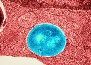
Staphylococcus aureus bacteria. Coloured transmission electron micrograph (TEM) of a Staphylococcus aureus bacterium (round) in skin tissue. S

Background imageBacterial Collection: Syphilis bacteria, artwork C016 / 8956

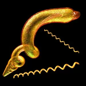
Syphilis bacteria, artwork C016 / 8956
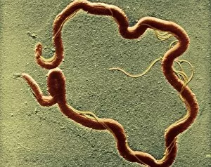
Syphilis bacteria. Computer artwork of the spirochaete bacteria Treponema pallidum, which cause syphilis. Syphilis is usually sexually transmitted

Background imageBacterial Collection: Shigella bacterium and macrophage, SEM C016 / 8922

Shigella bacterium and macrophage, SEM C016 / 8922
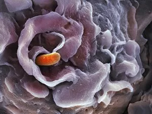
Shigella bacterium. Coloured scanning electron micrograph (SEM) of a Shigella sp. bacterium (orange) being engulfed by a macrophage white blood cell. This process is called phagocytosis

Background imageBacterial Collection: Mycobacterium fortuitum bacteria C016 / 9424

Mycobacterium fortuitum bacteria C016 / 9424
Mycobacterium fortuitum bacteria, coloured scanning electron micrograph (SEM). These bacteria (blue) are a non-tuberculosis strain of Mycobacterium

Background imageBacterial Collection: Orientia tsutsugamushi bacterium C016 / 9431

Orientia tsutsugamushi bacterium C016 / 9431
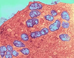
Orientia tsutsugamushi bacterium. Transmission electron micrograph (TEM) of a section through an Orientia tsutsugamushi bacterium. O

Background imageBacterial Collection: E. coli bacteria, SEM C016 / 9130

E. coli bacteria, SEM C016 / 9130
E. coli bacteria. Coloured scanning electron micrograph (SEM) of Escherichia coli bacteria (red) attached to the remains of a dead cell. E

Background imageBacterial Collection: Pseudomonas aeruginosa bacterium, SEM C016 / 9441

Pseudomonas aeruginosa bacterium, SEM C016 / 9441
Pseudomonas aeruginosa bacterium, coloured transmission electron micrograph (TEM). P. aeruginosa is a Gram-negative rod-shaped bacterium found in soil and water

Background imageBacterial Collection: Shigella bacterium and macrophage, SEM C016 / 8924

Shigella bacterium and macrophage, SEM C016 / 8924
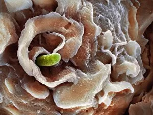
Shigella bacterium. Coloured scanning electron micrograph (SEM) of a Shigella sp. bacterium (green) being engulfed by a macrophage white blood cell. This process is called phagocytosis

Background imageBacterial Collection: MRSA bacteria, SEM C016 / 9422

MRSA bacteria, SEM C016 / 9422
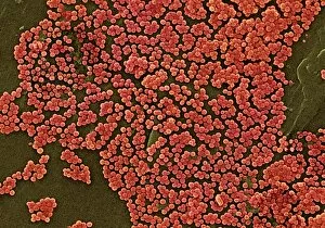
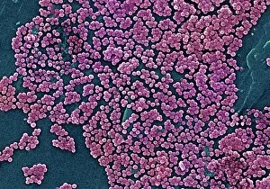
MRSA bacteria. Coloured scanning electron micrograph (SEM) of methicillin-resistant Staphylococcus aureus (MRSA, round) bacteria

Background imageBacterial Collection: Syphilis bacterium, TEM C016 / 9461

Syphilis bacterium, TEM C016 / 9461
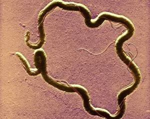
Syphilis bacteria. Transmission electron micrograph (TEM) of a Treponema pallidum spirochaete bacterium. T. pallidum is the bacteria that is responsible for the sexually transmitted infection syphilis

Background imageBacterial Collection: E. coli bacteria, SEM C016 / 9127

E. coli bacteria, SEM C016 / 9127
E. coli bacteria. Coloured scanning electron micrograph (SEM) of Escherichia coli bacteria (green) attached to the remains of a dead cell. E

Background imageBacterial Collection: Helicobacter pylori bacteria, SEM C016 / 9135

Helicobacter pylori bacteria, SEM C016 / 9135
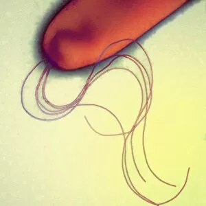
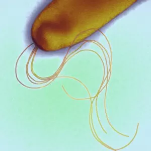
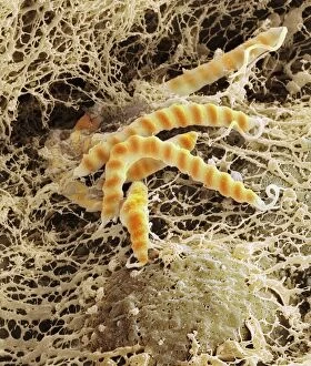
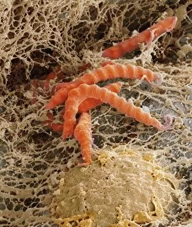
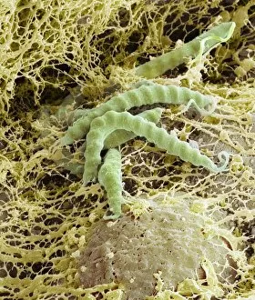
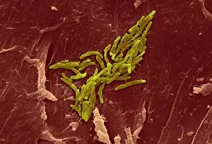
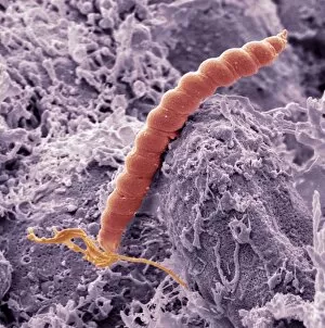
Helicobacter pylori bacteria. Coloured scanning electron micrograph (SEM) of Helicobacter pylori bacteria (yellow) in the stomach. H

Background imageBacterial Collection: Helicobacter pylori bacteria, SEM C016 / 9134

Helicobacter pylori bacteria, SEM C016 / 9134
Helicobacter pylori bacteria. Coloured scanning electron micrograph (SEM) of Helicobacter pylori bacteria (orange) in the stomach. H

Background imageBacterial Collection: Shigella bacterium and macrophage, SEM C016 / 8923

Shigella bacterium and macrophage, SEM C016 / 8923
Shigella bacterium. Coloured scanning electron micrograph (SEM) of a Shigella sp. bacterium (orange) being engulfed by a macrophage white blood cell. This process is called phagocytosis

Background imageBacterial Collection: Helicobacter pylori bacteria, SEM C016 / 9136

Helicobacter pylori bacteria, SEM C016 / 9136
Helicobacter pylori bacteria. Coloured scanning electron micrograph (SEM) of Helicobacter pylori bacteria (green) in the stomach. H

Background imageBacterial Collection: Gram-stained bacteria C016 / 7500

Gram-stained bacteria C016 / 7500
Gram-stained bacteria. Computer artwork showing gram-positive (blue) and gram-negative (red) bacteria. Gram-positive bacteria are those that are stained dark blue or violet by Gram staining

Background imageBacterial Collection: Syphilis bacterium, TEM C016 / 9462

Syphilis bacterium, TEM C016 / 9462
Syphilis bacteria. Transmission electron micrograph (TEM) of a Treponema pallidum spirochaete bacterium. T. pallidum is the bacteria that is responsible for the sexually transmitted infection known

Background imageBacterial Collection: Staphylococcus aureus bacteria, TEM C016 / 9457

Staphylococcus aureus bacteria, TEM C016 / 9457
Staphylococcus aureus bacteria. Coloured transmission electron micrograph (TEM) of a Staphylococcus aureus bacterium (blue) in skin tissue. S

Background imageBacterial Collection: Mycobacterium fortuitum bacteria C016 / 9423

Mycobacterium fortuitum bacteria C016 / 9423
Mycobacterium fortuitum bacteria, coloured scanning electron micrograph (SEM). These bacteria (yellow) are a non-tuberculosis strain of Mycobacterium

Background imageBacterial Collection: Gonorrhoea bacteria, TEM C016 / 9425

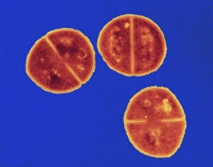
Gonorrhoea bacteria, TEM C016 / 9425
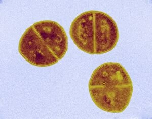
Gonorrhoea bacteria. Transmission electron micrograph (TEM) of a diplococcal pair of Neisseria gonorrhoeae bacteria. N. gonorrhoeae is the bacteria that is responsible for the sexually transmitted

Background imageBacterial Collection: Staphylococcus aureus bacteria, TEM C016 / 9455

Staphylococcus aureus bacteria, TEM C016 / 9455
Staphylococcus aureus bacteria. Coloured transmission electron micrograph (TEM) of Staphylococcus aureus bacteria four hours after infection of pulmonary epithelial cells. S

Background imageBacterial Collection: Orientia tsutsugamushi bacteria infection C016 / 9429

Orientia tsutsugamushi bacteria infection C016 / 9429
Orientia tsutsugamushi bacteria infection. Transmission electron micrograph (TEM) of a section through a capillary (red) infected with Orientia tsutsugamushi bacteria (blue). O

Background imageBacterial Collection: MRSA bacteria, SEM C016 / 9421

MRSA bacteria, SEM C016 / 9421
MRSA bacteria. Coloured scanning electron micrograph (SEM) of methicillin-resistant Staphylococcus aureus (MRSA, round) bacteria

Background imageBacterial Collection: Legionella infected protozoan, TEM C016 / 9412

Legionella infected protozoan, TEM C016 / 9412
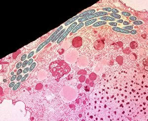
Legionella infected protozoan. Transmission electron micrograph (TEM) of a section through a single-celled Tetrahymena pyriformis protozoan, infected with Legionella pneumophila bacteria (yellow)

Background imageBacterial Collection: Orientia tsutsugamushi bacterium C016 / 9430

Orientia tsutsugamushi bacterium C016 / 9430
Orientia tsutsugamushi bacterium. Transmission electron micrograph (TEM) of a section through an Orientia tsutsugamushi bacterium. O

Background imageBacterial Collection: Helicobacter pylori bacteria, SEM C016 / 9053

Helicobacter pylori bacteria, SEM C016 / 9053
Helicobacter pylori bacteria. Coloured scanning electron micrograph (SEM) of a Helicobacter pylori bacterium (blue) in the stomach. H

Background imageBacterial Collection: Staphylococcus aureus bacteria, TEM C016 / 9456

Staphylococcus aureus bacteria, TEM C016 / 9456
Staphylococcus aureus bacteria. Coloured transmission electron micrograph (TEM) of Staphylococcus aureus bacteria four hours after infection of pulmonary epithelial cells. S

Background imageBacterial Collection: Meningitis bacteria, SEM

Meningitis bacteria, SEM
Meningitis bacteria. Coloured scanning electron micrograph (SEM) of Neisseria meningitidis bacteria. N. meningitidis is an obligate human parasite that causes meningococcal meningitis

Background imageBacterial Collection: Anthrax toxin prepore, molecular model

Anthrax toxin prepore, molecular model
Anthrax toxin prepore. Molecular model of protective antigen (PA) from anthrax (Bacillus anthracis) bacteria complexed with an anthrax toxin receptor, forming a prepore (hole, centre)

Background imageBacterial Collection: Bacterial protease molecule

Bacterial protease molecule. Computer model of a molecule of HsIUV protease complexed with its chaperone protein from a bacterium. Proteases are enzymes that break down proteins

Background imageBacterial Collection: Bacterial ribosome and protein synthesis

Bacterial ribosome and protein synthesis. Molecular model showing a bacterial ribosome reading an mRNA (messenger ribonucleic acid) strand (blue) and synthesising a protein

Background imageBacterial Collection: Stomach ulcer bacteria origins, artwork

Stomach ulcer bacteria origins, artwork
Stomach ulcer bacteria origins. Artwork of a human stomach and a magnified view of the Helicobacter pylori bacterium (green) that causes stomach ulcers


All Professionally Made to Order for Quick Shipping

"Bacterial: Unveiling the Microscopic World of Disease and Discovery" Step into a time capsule as we journey through history, exploring the captivating realm of bacteria. From the haunting presence of plague doctors in 17th-century artwork to cutting-edge scientific images captured by scanning electron microscopes (SEM), prepare to be fascinated by these tiny yet formidable organisms. Witness the battle between our immune system and Methicillin-resistant Staphylococcus aureus (MRSA) as a brave neutrophil engulfs it under SEM C018/8596. Marvel at the groundbreaking Calots spinal surgery from the 19th century, which revolutionized medical practices despite lurking bacterial threats. Peering through an X-ray, tuberculosis reveals its sinister grip on human lungs while skin disorders come alive in vivid artwork, reminding us of bacteria's impact on our bodies. E. coli bacteria, magnified under SEM, showcases their intricate structures that belie their harmful potential. Travel back to Soviet Russia in 1967 with a cholera vaccination poster that symbolizes humanity's fight against bacterial epidemics. Observe salmonella bacteria under SEM; their deceptively beautiful appearance belies their ability to cause severe illness. Delve into historical diagrams depicting anthrax cultures and learn about Yersinia pestis - responsible for devastating pandemics throughout history. The tuberculosis bacterium reminds us of its long-standing presence as one of humanity's greatest adversaries. Finally, witness nature's artistic flair with spiral spore chains formed by Streptomyces bacteria – showcasing both beauty and resilience within this microscopic world. Through these glimpses into bacterial realms past and present, we gain insight into our ongoing struggle against infectious diseases. Let this exploration ignite curiosity about microbial lifeforms that shape our existence – forever reminding us how knowledge can empower us in combating these invisible foes.