
Bacteriology Collection
"Bacteriology: Exploring the Microscopic World of Skin Disorders and Artwork" Delve into the fascinating realm of bacteriology


All Professionally Made to Order for Quick Shipping
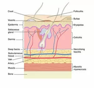
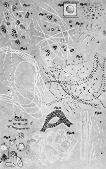
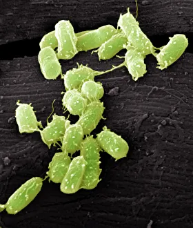
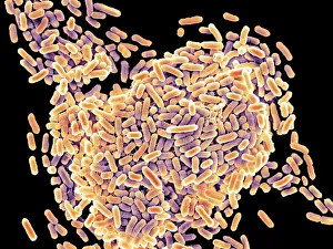
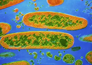
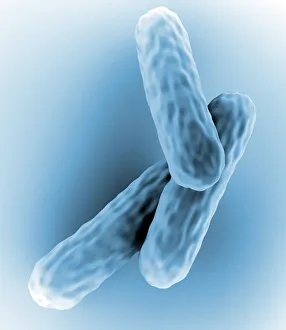
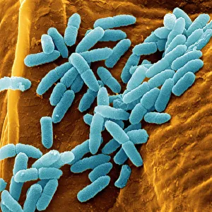
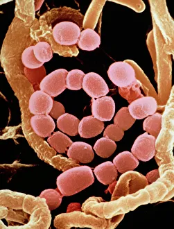
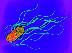
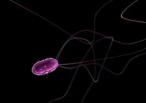
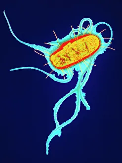
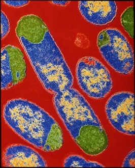
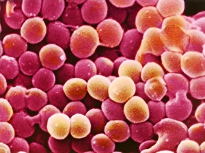
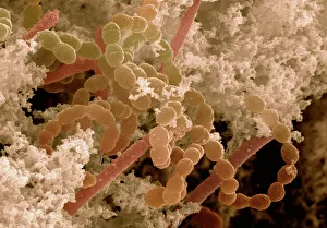
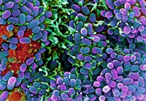
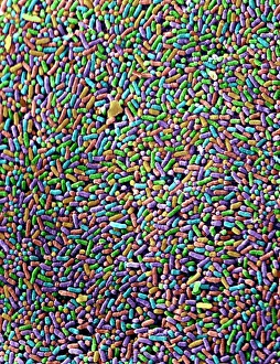
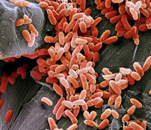
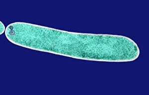
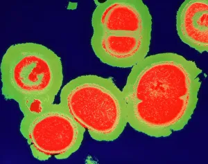
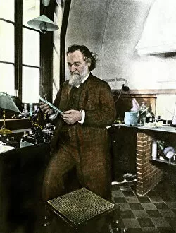
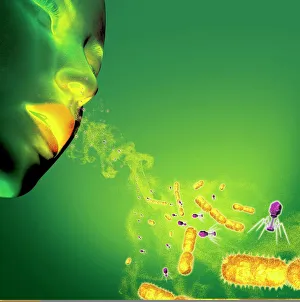
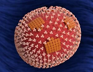
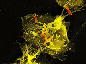
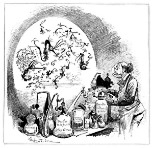
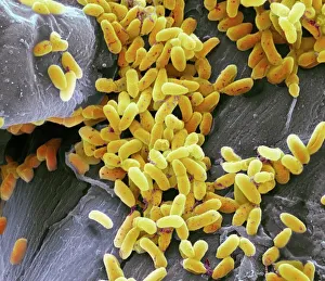
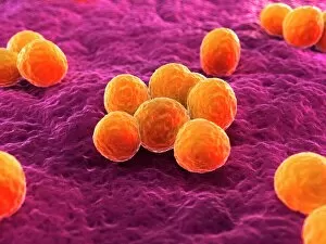
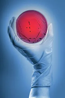
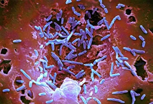
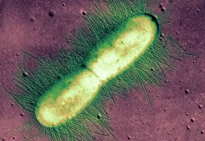
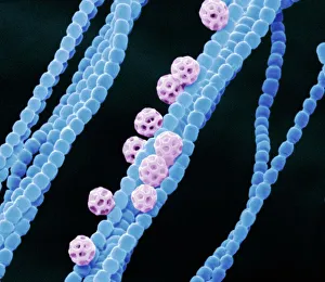
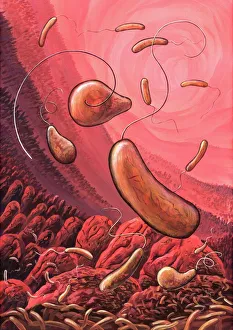
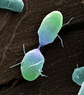
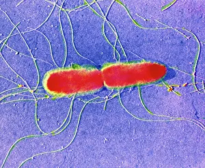
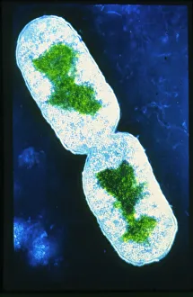
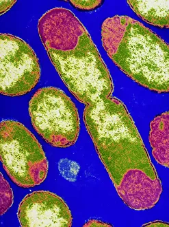
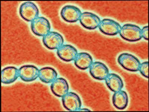
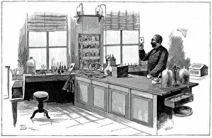
"Bacteriology: Exploring the Microscopic World of Skin Disorders and Artwork" Delve into the fascinating realm of bacteriology, where intricate artwork meets skin disorders. Through scanning electron microscopy (SEM), we witness the captivating beauty of E. Coli bacteria, their distinctive rod-like shapes forming a mesmerizing pattern. Salmonella bacteria also reveal their unique structure under SEM, showcasing their spherical forms with intriguing surface details. In a colored transmission electron micrograph (TEM), Yersinia pestis bacteria come to life in vibrant hues, highlighting the diversity within cell types. This artistic representation allows us to appreciate the complexity and intricacy of these microscopic organisms that can cause severe diseases like plague. Travel back in time as historical diagrams depict Anthrax cultures, revealing how scientists once studied this deadly disease. The tuberculosis bacteria are captured in all their glory; their slender rods painting a somber picture of one of humanity's oldest foes. Witness nature's creativity through the spiral spore chain formation of Streptomyces bacteria – an enchanting display resembling delicate strands woven together with precision. Flagellate bacteria showcase their remarkable motility through whip-like appendages called flagella, propelling themselves forward with grace and agility. Elevating our understanding further is an up-close encounter with Staphylococcus aureus bacteria – notorious for causing various infections ranging from minor skin conditions to life-threatening illnesses. Their distinct clusters become apparent as they thrive amidst human hosts. Amongst this diverse microbial world stands E. coli bacterium - its presence ubiquitous yet often misunderstood due to its association with foodborne illnesses. However, it plays crucial roles in digestion and research breakthroughs alike. Bacteriology unravels the hidden wonders within our microbiome while shedding light on pathogens that challenge human health daily. As we explore these captivating images and delve deeper into this field, we gain insights into both artistry at a microscopic level and the complex interplay between bacteria and human biology.