
Bio Chemistry Collection (page 2)
Biochemistry is the captivating realm where science and life intertwine, revealing the intricate secrets of our existence


All Professionally Made to Order for Quick Shipping
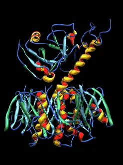
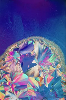
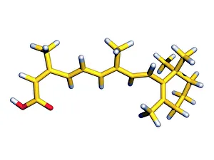
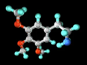
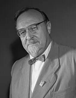
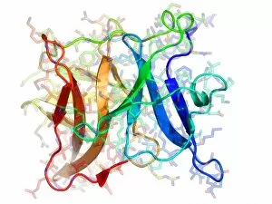
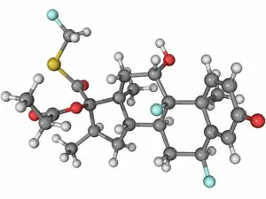
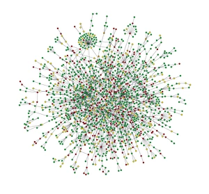
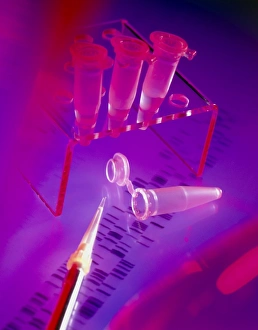
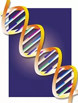
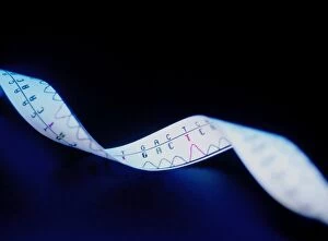
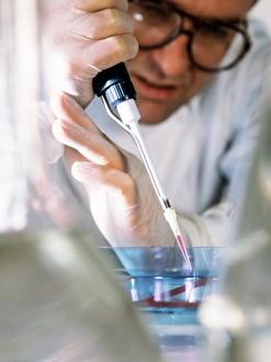
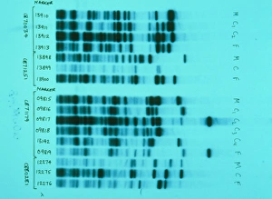
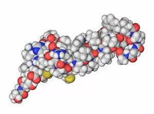
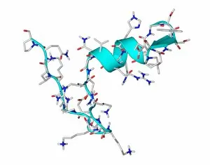
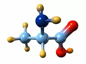
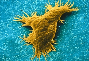
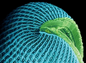
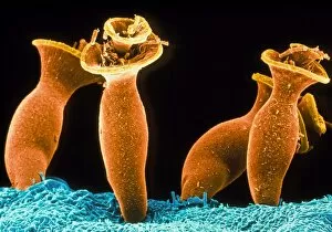
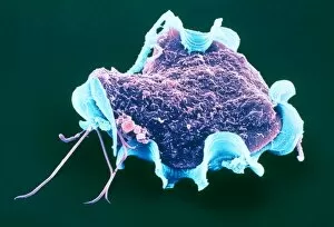
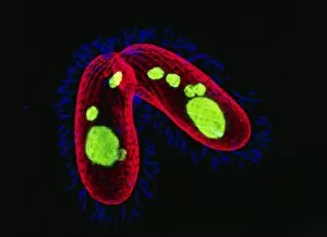
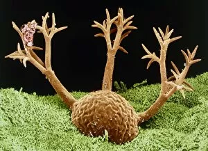
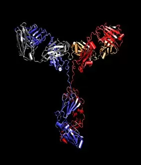
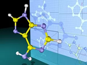
Biochemistry is the captivating realm where science and life intertwine, revealing the intricate secrets of our existence. As I gaze at the computer screen displaying a mesmerizing human genetic sequence, I am reminded of the boundless potential encoded within each double-stranded RNA molecule. The elegant dance of DNA transcription unfolds before my eyes, its molecular model illuminating the blueprint of life itself. In another corner, caffeine crystals come to life under a light micrograph, reminding us that even in our daily rituals lies an underlying biochemical symphony. The iconic DNA molecule stands tall as a symbol of discovery and progress, thanks to the pioneering work of Watson and Crick who unraveled its mysteries. Isaac Asimov's brilliance shines through as we acknowledge his contributions not only as a renowned US author but also as a biochemist who bridged literature with scientific exploration. Artistic renditions capture the beauty and complexity of metabolic enzymes and secondary structures of proteins, showcasing nature's ingenuity at every turn. The quest for knowledge extends into brain protein research; unlocking these enigmatic molecules could hold answers to understanding neurological disorders that plague humanity. A stunning computer artwork reveals beta DNA segments intertwined with spheres like celestial bodies orbiting their own gravitational pull - an awe-inspiring representation of interconnectedness on both macroscopic and microscopic scales. Amidst it all lies the nucleotide base matrix - an intricate web connecting all living beings across time and space. Biochemistry beckons us to explore this matrix further; deciphering its language holds profound implications for medicine, agriculture, biotechnology, and beyond. In this captivating world where science meets life's building blocks, biochemistry invites us to unravel nature's deepest secrets while inspiring wonderment at every step along this remarkable journey.