
Biological Collection (page 5)
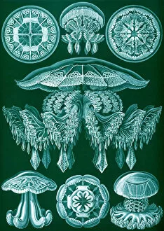
"Exploring the Intricate Beauty of Biology: From Garden Paintings to X-rays" Step into the mesmerizing world of biology as we unravel its captivating secrets


All Professionally Made to Order for Quick Shipping
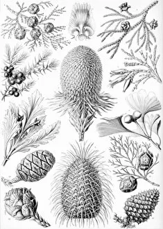
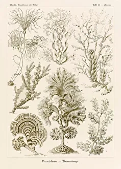
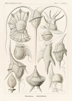
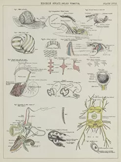
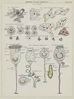
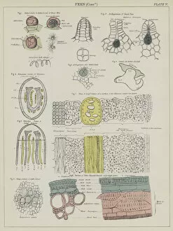
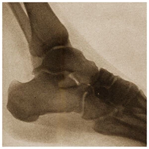
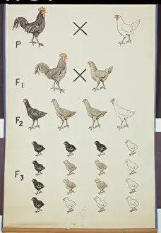
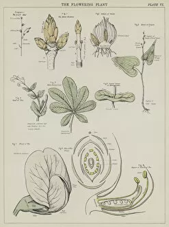
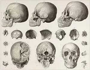
"Exploring the Intricate Beauty of Biology: From Garden Paintings to X-rays" Step into the mesmerizing world of biology as we unravel its captivating secrets. The Garden Paintings from the so-called Villa of Livia transport us to a time where nature's beauty was immortalized on canvas, showcasing the delicate balance between art and science. Delve deeper into the microscopic realm with Collagen, captured in stunning detail through Scanning Electron Micrograph (SEM). Witness its intricate structure, reminding us of the building blocks that hold our bodies together. Embark on a journey inside our minds as we observe Brain and brain waves during rest, beautifully illustrated. Discover the symphony of neural activity that occurs when we find solace in relaxation. Marvel at the enigmatic Praying Mantis head, an embodiment of predatory prowess. Its keen senses and formidable appearance serve as a testament to nature's ingenuity. Behold Pinus cembra, also known as Arolla pine, standing tall amidst rugged landscapes. This resilient tree symbolizes endurance and adaptability in even the harshest environments. Uncover the silent hero within our bodies –the Liver– tirelessly working behind-the-scenes to keep us healthy. Appreciate its vital role in detoxification and metabolism. Witness Amaryllis like never before through an X-ray lens; revealing hidden intricacies beneath its vibrant petals. Nature's artistry is not limited to what meets our eyes alone. Zooming out from microcosms to everyday objects, explore Ariel low temperature washing powder gel up close - Close up of plastic bottle UK reveals how innovation blends seamlessly with convenience for modern living. Peek into medical imaging technology with Leg in stiletto shoe MRI style - X-ray capturing both fashion statement and anatomy simultaneously. It reminds us that beauty can be found even within unexpected places. Admire Tulip (Tulipa sp. ) through an ethereal X-ray, where its delicate structure is unveiled.