
Blastocyst Collection
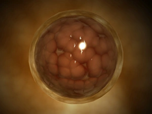
The human blastocyst, depicted in artwork F006/2190, is a crucial stage of early embryonic development


All Professionally Made to Order for Quick Shipping
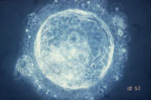
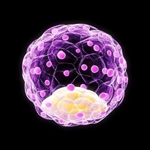
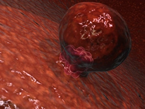
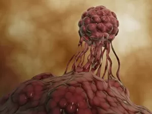
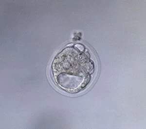
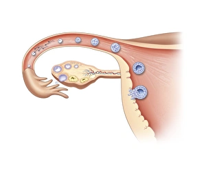
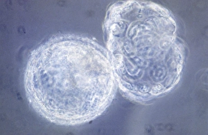
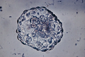
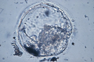
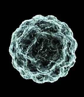
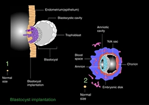
The human blastocyst, depicted in artwork F006/2190, is a crucial stage of early embryonic development. This tiny cluster of cells marks the beginning of life as it starts to implant itself into the wall of the uterus. With its microscopic view, we witness the awe-inspiring process of stem cell development within this blastocyst. During pregnancy, this blastocyst transforms into a blastula, as seen through the lens of a microscope in picture No. 10875811 and 10875812. The intricate details captured in these images showcase the complexity and beauty hidden within this minuscule structure. For those struggling with infertility or seeking alternative methods for conception, IVF treatment (depicted in F008/3570 and F008/3571) offers hope by assisting in creating viable blastocysts outside the body before transferring them to the uterus. As we delve deeper into understanding embryonic development, studying and observing these blastocysts provide invaluable insights that can potentially revolutionize reproductive medicine. From their formation to implantation, every step holds immense significance on our journey towards unraveling life's mysteries at its earliest stages. Whether marveling at an artwork or peering through a microscope lens at these remarkable structures like Blastocyst embryo (light micrograph F008 / 3573), one cannot help but be captivated by their potential and significance in shaping human existence.