Fraud Blocker Skip to main content

Blood Vessels Collection

Blood vessels are the intricate network of highways that carry life-sustaining blood throughout our bodies

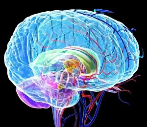
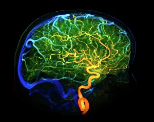
Background imageBlood Vessels Collection: Brain blood vessels, 3D angiogram C007 / 1981

Brain blood vessels, 3D angiogram C007 / 1981
Brain blood vessels. Coloured 3D lateral angiogram (blood vessel X-ray) of the blood vessels in the brain of a healthy 32-year-old

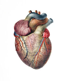
Background imageBlood Vessels Collection: Heart

Heart. Historical anatomical artwork of the human heart, seen from the front

Background imageBlood Vessels Collection: Brain anatomy, artwork

Brain anatomy, artwork
Brain anatomy, computer artwork. The cerebellum is purple the corpus callosum is green

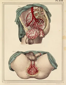
Background imageBlood Vessels Collection: Male groin arteries, 1825 artwork

Male groin arteries, 1825 artwork
Male groin arteries. Dissections of a male groin to show areas supplied by the internal iliac artery (red) and its branches

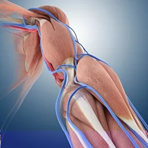
Background imageBlood Vessels Collection: Arm circulation, anatomical artwork C013 / 7419

Arm circulation, anatomical artwork C013 / 7419
Arm anatomy. Computer artwork showing the muscles (brown), ligaments (white), arteries (red), veins (blue), lymph vessels (yellow), and lymph nodes (round, yellow) of a human arm

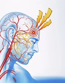
Background imageBlood Vessels Collection: Migraine pain

Migraine pain
Migraine. Conceptual artwork of lightning flashes marking the epicentre of pain in a mans forehead

Background imageBlood Vessels Collection: Pancreas anatomy, artwork

Pancreas anatomy, artwork
Pancreas anatomy. Cut-away computer artwork of the pancreas, which secretes digestive enzymes and the hormones insulin and glucagon, which regulate blood sugar levels

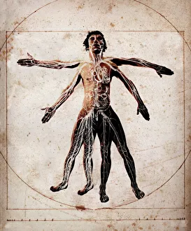
Background imageBlood Vessels Collection: Human anatomy

Human anatomy. Man with his limbs in two poses, overlaid with an artwork of his internal organs and blood vessels

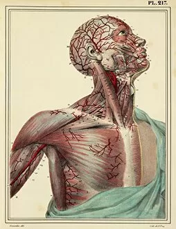
Background imageBlood Vessels Collection: Head and chest arteries, 1825 artwork

Head and chest arteries, 1825 artwork
Head and chest arteries. Superficial arteries (red) of the chest, neck and head, which are visible when the skin and muscle fascia are removed

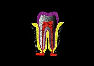
Background imageBlood Vessels Collection: Human tooth anatomy, artwork

Human tooth anatomy, artwork
Human tooth anatomy. Artwork of a cross-section through a human tooth to show its anatomical structure. The two main areas are the crown (above the gum-line) and the root (embedded in the gum)

Background imageBlood Vessels Collection: Mascagni artwork of human heart with its nerves

Mascagni artwork of human heart with its nerves
Human heart. Computer-enhanced historical artwork of the healthy human heart, with its blood vessels and nerves

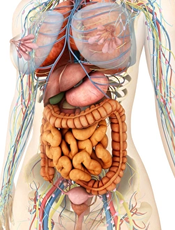
Background imageBlood Vessels Collection: Female body showing digestive and circulatory system

Female body showing digestive and circulatory system
Medical illustration of female body showing digestive and circulatory system, perspective view

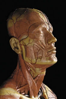
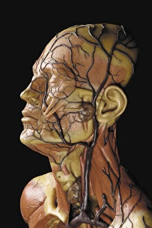
Background imageBlood Vessels Collection: Detail of a male anatomical ecorche model, displaying muscles, arteries and veins

Detail of a male anatomical ecorche model, displaying muscles, arteries and veins
CH469822 Detail of a male anatomical ecorche model, displaying muscles, arteries and veins, the circulation and nervous system (wax) (see also 469823 to 469827) by Zeiller

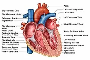
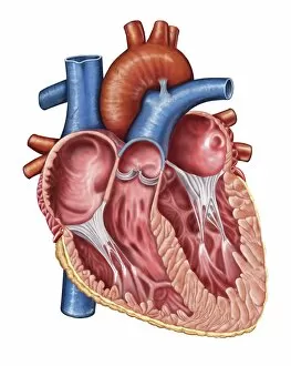
Background imageBlood Vessels Collection: Anatomy of heart interior, frontal section

Anatomy of heart interior, frontal section

Background imageBlood Vessels Collection: Perspective view of human body, whole organs and bones

Perspective view of human body, whole organs and bones

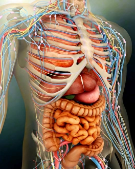
Background imageBlood Vessels Collection: Human body with internal organs, nervous system, lymphatic system and circulatory system

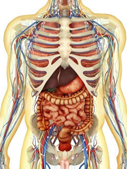
Human body with internal organs, nervous system, lymphatic system and circulatory system
Transparent human body with internal organs, nervous system, lymphatic system and circulatory system

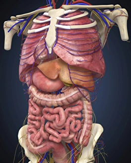
Background imageBlood Vessels Collection: Midsection view showing internal organs of human body

Midsection view showing internal organs of human body

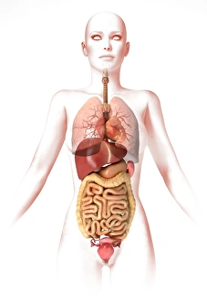
Background imageBlood Vessels Collection: Anatomy of female body with internal organs

Anatomy of female body with internal organs

Background imageBlood Vessels Collection: Male chest anatomy of thorax with heart, veins, arteries and lungs

Male chest anatomy of thorax with heart, veins, arteries and lungs

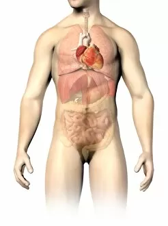
Background imageBlood Vessels Collection: Male anatomy of internal organs

Male anatomy of internal organs, with heart highlighted and rest of organs in half tone

Background imageBlood Vessels Collection: Gross anatomy of the human heart

Gross anatomy of the human heart

Background imageBlood Vessels Collection: Screws for compression of blood vessels by Jean Louis Petite

Screws for compression of blood vessels by Jean Louis Petite
FLO4611834 Screws for compression of blood vessels by Jean Louis Petite, tourniquet device and double band to adjust it, chamois skin compress

Background imageBlood Vessels Collection: Venous blood in arterial blood transformation in the lungs cell

Venous blood in arterial blood transformation in the lungs cell
7295211 Venous blood in arterial blood transformation in the lungs cell. Above: end of a bronchial branching

Background imageBlood Vessels Collection: Yoga roots

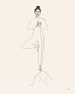
Yoga roots
Amanda Leon

Background imageBlood Vessels Collection: Metamorphosis

Metamorphosis
Amanda Leon

Background imageBlood Vessels Collection: Yellow ground, red heart

Yellow ground, red heart
E. de Juan

Background imageBlood Vessels Collection: Muscles and circulation of the head and neck, 1822-26. Creator: William Home Lizars

Muscles and circulation of the head and neck, 1822-26. Creator: William Home Lizars
Muscles and circulation of the head and neck, 1822-26. Plate IX from A System of Anatomical Plates of the Human Body, vol. 2

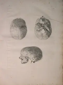
Background imageBlood Vessels Collection: Albinus II, Pl. III, Cranial bone, illustration from Tabulae ossium humanorum

Albinus II, Pl. III, Cranial bone, illustration from Tabulae ossium humanorum
HMB306101 Albinus II, Pl. III, Cranial bone, illustration from Tabulae ossium humanorum, by Bernhard Siegfried Albinus (1697-1770), published by J.&H. Verbeek, bibliop

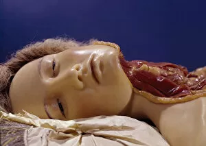
Background imageBlood Vessels Collection: Model of a man whose throat has been cut (wax)

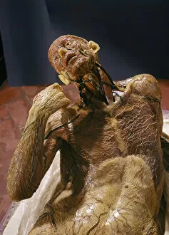
Model of a man whose throat has been cut (wax)
BEN360184 Model of a man whose throat has been cut (wax) by Susini, Clemente (1754-1814); Museo della Specola, Florence

Background imageBlood Vessels Collection: Model of a man whose throat has been cut (wax)

Model of a man whose throat has been cut (wax)
BEN360183 Model of a man whose throat has been cut (wax) by Susini, Clemente (1754-1814); Museo della Specola, Florence

Background imageBlood Vessels Collection: Model of a skinless man (wax)

Model of a skinless man (wax)
BEN360194 Model of a skinless man (wax) by Susini, Clemente (1754-1814); Museo della Specola, Florence

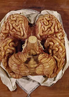
Background imageBlood Vessels Collection: Model of a brain (wax)

Model of a brain (wax)
BEN360191 Model of a brain (wax) by Susini, Clemente (1754-1814); Museo della Specola, Florence, Italy; (add.info.: cervello; scultura di cera; ); Photo eRaffaello Bencini; Italian, out of copyright

Background imageBlood Vessels Collection: Skull and Blood Vessels of the Human Head, 1913 (chromolithograph)

Skull and Blood Vessels of the Human Head, 1913 (chromolithograph)
2622111 Skull and Blood Vessels of the Human Head, 1913 (chromolithograph) by American School, (20th century); Private Collection; (add.info.: Vintage anatomical study of the male human head)

Background imageBlood Vessels Collection: Detail of a male anatomical ecorche model, displaying muscles, arteries and veins

Detail of a male anatomical ecorche model, displaying muscles, arteries and veins
CH469823 Detail of a male anatomical ecorche model, displaying muscles, arteries and veins, the circulation and nervous system (wax) (see also 469822 to 469827) by Zeiller

Background imageBlood Vessels Collection: Model of a skinless man (wax)

Model of a skinless man (wax)
BEN360193 Model of a skinless man (wax) by Susini, Clemente (1754-1814); Museo della Specola, Florence

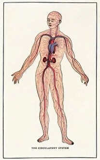
Background imageBlood Vessels Collection: Vintage Anatomical Print of the Human Circulatory System, 1912 (screen print)

Vintage Anatomical Print of the Human Circulatory System, 1912 (screen print)
3034946 Vintage Anatomical Print of the Human Circulatory System, 1912 (screen print) by American School

Background imageBlood Vessels Collection: Heart on a Noose

Heart on a Noose
Petri Damstén

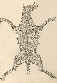
Background imageBlood Vessels Collection: Anatomia del Cavallo. Infirmita del Cavallo, 1602. Creator: Carlo Ruini

Anatomia del Cavallo. Infirmita del Cavallo, 1602. Creator: Carlo Ruini
Anatomia del Cavallo. Infirmita del Cavallo, 1602

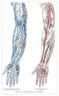
Background imageBlood Vessels Collection: Arms Blood vessels anatomy engraving 1857

Arms Blood vessels anatomy engraving 1857
Rank, johannes - The human being. 1 - 1894

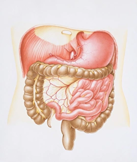
Background imageBlood Vessels Collection: Diagram of the abdominal cavity in the human body with the liver removed

Diagram of the abdominal cavity in the human body with the liver removed

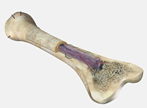
Background imageBlood Vessels Collection: Human bone, cross section diagram of Femur showing osteon, veins, marrow

Human bone, cross section diagram of Femur showing osteon, veins, marrow

Background imageBlood Vessels Collection: Illustration of yoke sac and amniotic membrane surrounding chicken embryo

Illustration of yoke sac and amniotic membrane surrounding chicken embryo

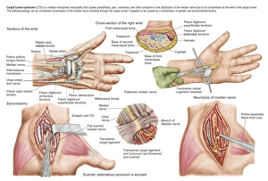
Background imageBlood Vessels Collection: Medical illustration showing carpal tunnel syndrome in the human wrist

Medical illustration showing carpal tunnel syndrome in the human wrist, and the surgical procedures associated with it

Background imageBlood Vessels Collection: Male human circulatory system, upper body

Male human circulatory system, upper body
Male human circulatory system, upper part, superimposed to realistic body

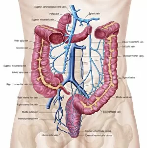
Background imageBlood Vessels Collection: Anatomy of human abdominal vein system

Anatomy of human abdominal vein system

Background imageBlood Vessels Collection: Interior of human heart

Interior of human heart

Background imageBlood Vessels Collection: Anatomy of human circulatory system

Anatomy of human circulatory system

Background imageBlood Vessels Collection: Human midsection with internal organs

Human midsection with internal organs

Background imageBlood Vessels Collection: Anatomy of female body with internal organs superimposed

Anatomy of female body with internal organs superimposed

Background imageBlood Vessels Collection: Human body showing heart and main circulatory system position

Human body showing heart and main circulatory system position
Transparent human body showing heart and main circulatory system position with internal organs, nervous system, lymphatic system and circulatory system

Background imageBlood Vessels Collection: Histopathology and pathophysiology of diabetic food ulcers

Histopathology and pathophysiology of diabetic food ulcers

Background imageBlood Vessels Collection: Female skeleton and organs, artwork

Female skeleton and organs, artwork
Female skeleton and organs and blood vessels, artwork

Background imageBlood Vessels Collection: Heart of stone

Heart of stone, computer artwork. The heart is a hollow sac (lower frame) of muscular tissue that pumps blood around the body. Small surface coronary blood vessels supply the heart muscle with blood

Background imageBlood Vessels Collection: Diagram illustrating the two layers of human skin, epidermis, dermis and hair follicle

Diagram illustrating the two layers of human skin, epidermis, dermis and hair follicle

Background imageBlood Vessels Collection: Diagram of the muscles and tendons in the human ankle and foot

Diagram of the muscles and tendons in the human ankle and foot

Background imageBlood Vessels Collection: Cross section of human heart

Cross section of human heart and, bottom left, coronary system with aorta, coronary arteries, coronary vein and blood vessels

Background imageBlood Vessels Collection: Human upper body showing bones, muscles and circulatory system

Human upper body showing bones, muscles and circulatory system

Background imageBlood Vessels Collection: Nephron detail, the functional unit of excretion in the human kidney

Nephron detail, the functional unit of excretion in the human kidney

Background imageBlood Vessels Collection: Anatomy of human heart, cross section

Anatomy of human heart, cross section with detailed internal structure

Background imageBlood Vessels Collection: Retina with macular degeneration

Retina with macular degeneration

Background imageBlood Vessels Collection: Anatomy of back of human hand

Anatomy of back of human hand

Background imageBlood Vessels Collection: Acute coronary syndrome - microvascular obstruction

Acute coronary syndrome - microvascular obstruction

Background imageBlood Vessels Collection: Cross section of human heart

Cross section of human heart

Background imageBlood Vessels Collection: Pulmonary embolism, pathway of embolus to the heart and lungs

Pulmonary embolism, pathway of embolus to the heart and lungs

Background imageBlood Vessels Collection: Anatomy of human kidney function

Anatomy of human kidney function

Background imageBlood Vessels Collection: Anterior view of the human heart

Anterior view of the human heart

Background imageBlood Vessels Collection: Human circulatory system

Human circulatory system

Background imageBlood Vessels Collection: Arteries and veins of the human body

Arteries and veins of the human body

Background imageBlood Vessels Collection: Anatomy of male urinary system

Anatomy of male urinary system

Background imageBlood Vessels Collection: Anatomy of human body and circulatory system, front view

Anatomy of human body and circulatory system, front view

Background imageBlood Vessels Collection: Renal glomerulus

Renal glomerulus
The renal glomerulus is a network (tuft) of capillaries that performs the first step of filtering blood

Background imageBlood Vessels Collection: Anatomy of bowmans glomerular capsule

Anatomy of bowmans glomerular capsule

Background imageBlood Vessels Collection: Early stage of kidney cancer with a tumor visible on the kidney

Early stage of kidney cancer with a tumor visible on the kidney

Background imageBlood Vessels Collection: Artist depcition of abdominal aortic aneuryism (with labels)

Artist depcition of abdominal aortic aneuryism (with labels)

Background imageBlood Vessels Collection: Diagram of a nephron, the functional unit of excretion in the kidney

Diagram of a nephron, the functional unit of excretion in the kidney
Diagram of a nephron, the functional unit of excretion in the human kidney

Background imageBlood Vessels Collection: Medical illustration of male chest with arteries, veins, heart and rib cage

Medical illustration of male chest with arteries, veins, heart and rib cage
Three dimensional medical illustration of male chest showing arteries, veins, heart and rib cage

Background imageBlood Vessels Collection: Face and neck nerves, 1825 artwork

Face and neck nerves, 1825 artwork
Face and neck nerves. Head and neck dissected to show the superficial nerves (white)

Background imageBlood Vessels Collection: Heart chamber anatomy, artwork C014 / 2029

Heart chamber anatomy, artwork C014 / 2029
Heart chamber anatomy. Cutaway artwork revealing the four internal heart chambers and the heart valves and tendons (white). The two upper chambers are the atria

Background imageBlood Vessels Collection: Thymus gland, light micrograph C015 / 4970

Thymus gland, light micrograph C015 / 4970
Thymus gland. Light micrograph of a section through tissue from the thymus gland, part of the lymphoid system. The dense areas are the thymic cortex, which produces lots of lymphocytes

Background imageBlood Vessels Collection: Right shoulder and nerve plexus, artwork C016 / 6811

Right shoulder and nerve plexus, artwork C016 / 6811
Right shoulder and nerve plexus. Artwork of the nerves (yellow), arteries (red), and bones of the right shoulder, seen from the front

Background imageBlood Vessels Collection: External carotid artery, 3D CT scan C016 / 6341

External carotid artery, 3D CT scan C016 / 6341
External carotid artery. Coloured 3D computed tomography (CT) angiogram (blood vessel scan) of the head showing a normal external carotid artery (red, in neck) and its branches

Background imageBlood Vessels Collection: Retina layers, SEM C018 / 0520

Retina layers, SEM C018 / 0520
Retina layers, coloured scanning electron micrograph (SEM). The retina of the human eye is found on the inside of the eyeball and is formed from a number of layers


All products are expertly crafted, using premium materials, tailored to your specifications and promptly shipped

Blood vessels are the intricate network of highways that carry life-sustaining blood throughout our bodies. From the brain to the heart, from the retina to the neck and shoulder arteries, they ensure that every organ receives vital nutrients and oxygen. The 3D angiogram C007 / 1981 reveals a mesmerizing view of brain blood vessels, showcasing their complexity and importance. The heart, often referred to as the powerhouse of our body, relies on a vast system to pump oxygenated blood to every corner. Meanwhile, X-ray images capture the intricacies of neck and shoulder arteries, reminding us how crucial these pathways are for maintaining overall health. Intriguingly detailed artwork showcases both brain anatomy and male groin arteries from an 1825 perspective. These depictions serve as a testament to humanity's long-standing fascination with understanding our own bodies. Anatomical artwork C013 / 7419 beautifully illustrates arm circulation while highlighting its significance in ensuring proper functioning. Similarly captivating is pancreas anatomy depicted in yet another stunning piece of artistry. Head and chest arteries portrayed in an 1825 artwork remind us that even centuries ago, humans recognized the importance of studying these essential conduits for sustaining life. Migraine pain serves as a stark reminder that when something goes awry within our intricate vascular system, they can have debilitating consequences. These glimpses into human anatomy showcase not only scientific knowledge but also artistic expression at its finest. Finally, abdominal arteries captured through X-ray P206 / 0309 provide insight into yet another complex region where blood vessels play a critical role in maintaining optimal health. Blood vessels truly embody the essence of human existence - connecting each part like threads woven together in an intricate tapestry. Understanding their structure and function allows us to appreciate just how remarkable our bodies are; delicate yet resilient systems working tirelessly behind-the-scenes to keep us alive and thriving.

© Copyright Media Storehouse, All Rights Reserved 2003 - 2024
Facebook     Pinterest     100% Payment Secure     Reviews IO