
Bowels Collection
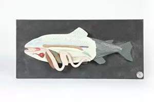
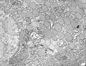
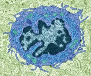
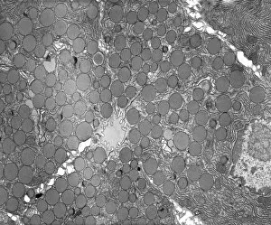
"Bowels: Unveiling the Intricate Depths of Anatomy and History" Delving into the depths of fish anatomy, we uncover a world hidden beneath shimmering scales


All Professionally Made to Order for Quick Shipping
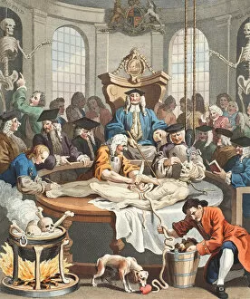
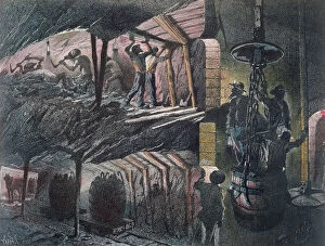
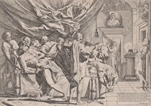
"Bowels: Unveiling the Intricate Depths of Anatomy and History" Delving into the depths of fish anatomy, we uncover a world hidden beneath shimmering scales. From gills to fins, their bowels hold secrets that unravel the mysteries of aquatic life. Intriguingly, history unveils a different kind of model - one stained with cruelty. The Horrible Cruelty of the Huguenots in France engraving depicts a dark chapter where human nature took an abhorrent turn. The Reward of Cruelty series by William Hogarth transports us to 18th-century England, showcasing etchings and engravings that expose mankind's capacity for brutality. Hand-colored or black-and-white photos capture these chilling scenes forever etched in our collective memory. Venturing further underground, we encounter a cross-section of a coal mine captured by Jules Ferat's skillful hand in vibrant litho colors. This visual representation reminds us not only of industry but also the sacrifices made within those treacherous tunnels. Yet amidst this darkness, there are glimpses of artistry intertwined with morbidity. A wax model depicting a disemboweled woman stands as both macabre curiosity and testament to anatomical study's intricate beauty. Shifting gears from physicality to conflict, an engraving titled "The War in the East" takes us back centuries ago when Shakespearean drama collided with Russian reality on Ibraila's stage. A sketch frozen in time captures this intriguing fusion between literature and geopolitics. Ancient rituals come alive through marble sculptures portraying haruspices consulting bull entrails - divination practices steeped in mysticism and belief systems long past their prime yet fascinating nonetheless. Artistic expressions take unexpected forms as well; oil on cradled wood panel presents "Evisceration of a Roebuck with a Portrait of Married Couple.