Fraud Blocker Skip to main content

Cell Body Collection

The cell body, also known as the soma or perikaryon, is a vital component of nerve cells

Background imageCell Body Collection: Nerve cell, SEM

Nerve cell, SEM
Nerve cell. Coloured scanning electron micrograph (SEM) of a nerve cell (neuron). Neurons are responsible for passing information around the central nervous system (CNS)

Background imageCell Body Collection: Nerve cell

Nerve cell. Computer artwork of a nerve cell, also called a neuron. Neurons are responsible for passing information around the central nervous system (CNS) and from the CNS to the rest of the body

Background imageCell Body Collection: Brain neuron

Brain neuron. Computer reconstruction of a medium spiny neuron from the basal ganglia of the brain. Neurons (nerve cells) are responsible for passing information around the central nervous system

Background imageCell Body Collection: Nerve cells, SEM

Nerve cells, SEM
Nerve cells. Coloured scanning electron micrograph (SEM) of nerve cells, known as neurones. Nerve cells occur in the brain, spinal cord, and in ganglia

Background imageCell Body Collection: Nerve cells, abstract artwork

Nerve cells, abstract artwork
Nerve cells. Abstract computer artwork of nerve cells, or neurons. Neurons are responsible for passing information around the central nervous system (CNS) and from the CNS to the rest of the body

Background imageCell Body Collection: Purkinje nerve cell, TEM C014 / 0583

Purkinje nerve cell, TEM C014 / 0583
Purkinje nerve cell. Transmission electron micrograph (TEM) of a purkinje nerve cell (green) from the cerebellum of the brain, showing the cell body (centre) and its primary dendrite (cell process)

Background imageCell Body Collection: Nerve cells, computer artwork

Nerve cells, computer artwork
Nerve cells. Computer artwork of nerve cells, or neurons

Background imageCell Body Collection: Microscopic view of a unipolar neuron

Microscopic view of a unipolar neuron. A unipolar neuron is a type of neuron in which only one protoplasmic process (neurite) extends from the cell body

Background imageCell Body Collection: Anatomy structure of neurons

Anatomy structure of neurons

Background imageCell Body Collection: Schematic of the hypothalamus receiving nerve impulses from the body

Schematic of the hypothalamus receiving nerve impulses from the body and sending messages to the circulatory and nervous system

Background imageCell Body Collection: Renal Corpuscle and the filtration membrane

Renal Corpuscle and the filtration membrane. Glomerular capillary surrounded by podocytes

Background imageCell Body Collection: Nerve with myelin sheath, seen in lower right, connects with muscle

Nerve with myelin sheath, seen in lower right, connects with muscle. Blood vessel and immune cells are seen in the center and upper right of image

Background imageCell Body Collection: Energy meridians of the human body

Energy meridians of the human body and a cell showing energy vibrational communication

Background imageCell Body Collection: Neurofibromatosis, a genetic disorder of the nervous system

Neurofibromatosis, a genetic disorder of the nervous system
A genetic disorder of the nervous system, neurofibromatosis causes tumors to form on nerves throughout the body, including a type of tumor called an optic nerve glioma that can result in childhood

Background imageCell Body Collection: Nerve cells, artwork F007 / 5512

Nerve cells, artwork F007 / 5512
Computer artwork of nerve cells, also called neurons. Neurons are responsible for passing information around the central nervous system (CNS) and from the CNS to the rest of the body

Background imageCell Body Collection: Nerve cell, artwork F005 / 6921

Nerve cell, artwork F005 / 6921
Computer artwork of a nerve cell, also called a neuron. Neurons are responsible for passing information around the central nervous system (CNS) and from the CNS to the rest of the body

Background imageCell Body Collection: Nerve cell, artwork F005 / 6919

Nerve cell, artwork F005 / 6919
Computer artwork of a nerve cell, also called a neuron. Neurons are responsible for passing information around the central nervous system (CNS) and from the CNS to the rest of the body

Background imageCell Body Collection: Nerve cells, computer artwork F007 / 1988

Nerve cells, computer artwork F007 / 1988
Nerve cells. Computer artwork of nerve cells, or neurons

Background imageCell Body Collection: Nerve synapse, computer artwork C017 / 3429

Nerve synapse, computer artwork C017 / 3429
Nerve synapse. Conceptual computer artwork of a synapse between two nerve cells or neurons. Neurons are responsible for passing information around the central nervous system (CNS)

Background imageCell Body Collection: Nerve cells, artwork C015 / 4265

Nerve cells, artwork C015 / 4265
Nerve cells. Computer artwork of two nerve cells or neurons. Neurons are responsible for passing information around the central nervous system (CNS) and from the CNS to the rest of the body

Background imageCell Body Collection: Nerve ganglion, light micrograph

Nerve ganglion, light micrograph
Nerve ganglion. Light micrograph of a section through a dorsal (sensory) spinal root ganglion associated with a sensory nerve root of the spinal cord. Sensory information from peripheral sites e.g

Background imageCell Body Collection: Myenteric nerve plexus, TEM

Myenteric nerve plexus, TEM
Myenteric nerve plexus. Transmission electron micrograph (TEM) of a section through a bundle of unmyelinated nerve fibres in the myenteric nerve plexus of the intestinal tract

Background imageCell Body Collection: Nerve cell, illustration C018 / 0740

Nerve cell, illustration C018 / 0740
Nerve cell, or neuron, illustration. Neurons are responsible for passing information around the central nervous system (CNS) and from the CNS to the rest of the body

Background imageCell Body Collection: Brain cells, artwork

Brain cells, artwork
Brain cells. Computer artwork representing neurons (nerve cells) in the brain

Background imageCell Body Collection: Purkinje nerve cell, TEM C014 / 0582

Purkinje nerve cell, TEM C014 / 0582
Purkinje nerve cell. Transmission electron micrograph (TEM) of a purkinje nerve cell (bright yellow, centre) from the cerebellum of the brain

Background imageCell Body Collection: Nerve cells, artwork C018 / 2887

Nerve cells, artwork C018 / 2887
Nerve cells, or neurons, computer artwork. Neurons are responsible for passing information around the central nervous system (CNS) and from the CNS to the rest of the body

Background imageCell Body Collection: Neural network, conceptual image C013 / 9958

Neural network, conceptual image C013 / 9958
Neural network, conceptual image. Computer artwork representing interconnecting nerve cells (neurons)

Background imageCell Body Collection: Neural network, conceptual image C013 / 9957

Neural network, conceptual image C013 / 9957
Neural network, conceptual image. Computer artwork representing interconnecting nerve cells (neurons)

Background imageCell Body Collection: Nerve cell, SEM C013 / 9772

Nerve cell, SEM C013 / 9772
Nerve cell. Coloured scanning electron micrograph (SEM) of a nerve cell (neuron). Neurons are responsible for passing information around the central nervous system (CNS)

Background imageCell Body Collection: Purkinje nerve cell C013 / 9763

Purkinje nerve cell C013 / 9763
Purkinje nerve cell. Light micrograph of a purkinje nerve cell (orange, centre) from the cerebellum of the brain. The cell comprises a flask-shaped cell body from which numerous processes (dendrites)

Background imageCell Body Collection: Purkinje nerve cells C013 / 9745

Purkinje nerve cells C013 / 9745
Purkinje nerve cells. Light micrograph of three purkinje nerve cells (across bottom) from the cerebellum of the brain. The cells comprise a flask-shaped cell body from which numerous processes

Background imageCell Body Collection: Motor neurone nerve cell and synapses

Motor neurone nerve cell and synapses
Motor neurone and synapses. Computer graphic of a motor neurone and synapses (glowing yellow bulges, at lower centre). The cell body is at centre

Background imageCell Body Collection: Computer graphic of a motor neuron nerve cell

Computer graphic of a motor neuron nerve cell
Motor neurone. Computer graphic of a motor neuron nerve cell. At centre is the cell body; at the centre of this is a red circle which is the cell nucleus

Background imageCell Body Collection: LM of nerve cells and fibres in brain tissue

LM of nerve cells and fibres in brain tissue
Brain tissue. Light micrograph of a section through the grey matter of a normal brain. There are several nerve cell bodies (objects in white areas)

Background imageCell Body Collection: Light micrograph of a nerve cell

Light micrograph of a nerve cell
Neuron. Scanning electron micrograph of a nerve cell with a branching axon. On the right is the cell body, which contains the nucleus

Background imageCell Body Collection: Coloured SEM of spinal cord nerve cells

Coloured SEM of spinal cord nerve cells

Background imageCell Body Collection: Nerve cells, light micrograph

Nerve cells, light micrograph
Nerve cells, or neurons, light micrograph. Neurons are responsible for passing information around the central nervous system (CNS) and from the CNS to the rest of the body

Background imageCell Body Collection: Purkinje nerve cell, light micrograph

Purkinje nerve cell, light micrograph
Purkinje nerve cell. Confocal light micrograph of a purkinje cell (stained with green fluorescent protein), a type of neuron (nerve cell)

Background imageCell Body Collection: Nerve cell, abstract artwork

Nerve cell, abstract artwork
Nerve cell. Abstract computer artwork of a nerve cell, or neuron. Neurons are responsible for passing information around the central nervous system (CNS) and from the CNS to the rest of the body

Background imageCell Body Collection: Foetal neurons

Foetal neurons. Light micrograph of human foetal neurons (nerve cells). Neurons are responsible for passing information around the central nervous system (CNS)

Background imageCell Body Collection: Alzheimers disease brain cell, TEM

Alzheimers disease brain cell, TEM
Alzheimers disease brain cell. Coloured transmission electron micrograph (TEM) of a neurofibrillary tangle in a nerve cell from the brain of a patient with Alzheimers disease


All Professionally Made to Order for Quick Shipping

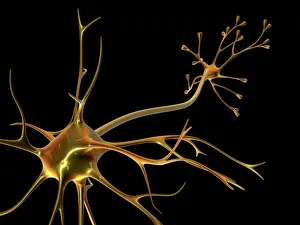
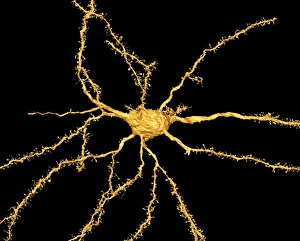
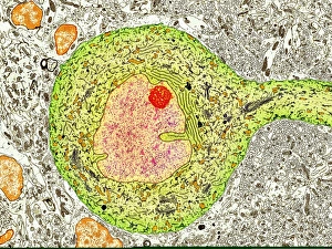
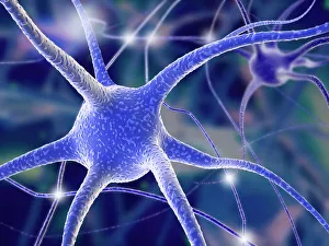
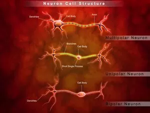
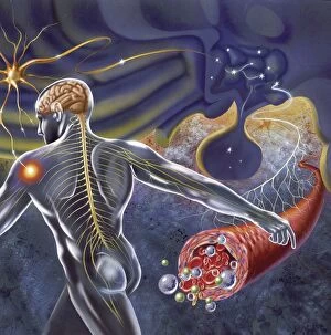
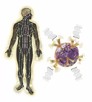
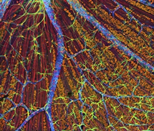
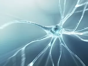
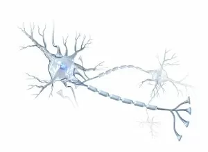
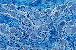
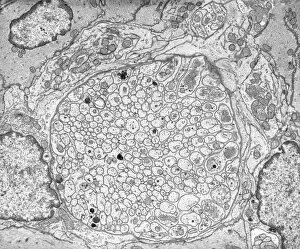
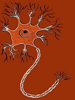
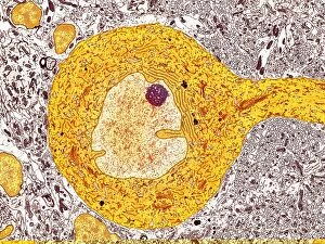
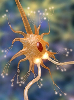
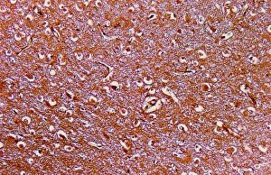
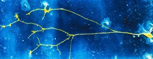
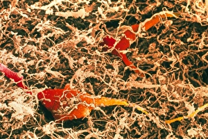
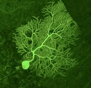
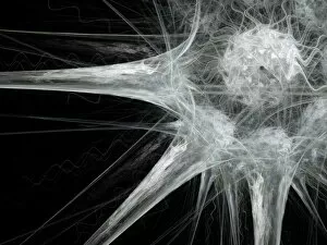
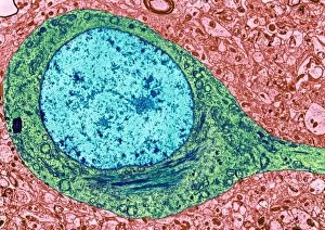
The cell body, also known as the soma or perikaryon, is a vital component of nerve cells. It serves as the control center and houses the nucleus, which contains genetic material necessary for cellular functions. In this captivating SEM image, we can observe the intricate details of a nerve cell's structure. Nerve cells, also called neurons, are specialized cells that transmit electrical signals throughout our bodies. This stunning SEM image showcases a network of interconnected nerve cells in all their glory. In this mesmerizing abstract artwork inspired by nerve cells, vibrant colors blend together to depict the complexity and beauty of these essential components of our nervous system. Using TEM technology (Transmission Electron Microscopy), scientists have captured an up-close view of a Purkinje nerve cell. The detailed image reveals its unique shape and internal structures with astonishing clarity. This computer-generated artwork provides us with an imaginative representation of how nerve cells might appear in a digital realm. The fusion of artistry and science offers us a glimpse into the fascinating world within our own bodies. Through light micrography techniques, researchers have captured this striking image showcasing numerous interconnected nerve cells. The delicate branches extend like tree roots, forming intricate networks that enable communication between different parts of our body. Art has always been inspired by nature's wonders; here we see an artist's interpretation capturing the essence and beauty of a single nerve cell through their creative lens. A microscopic view unveils the intriguing structure of a unipolar neuron - one type among many found in our nervous system. Its elongated shape allows it to efficiently transmit signals over long distances within our body. Understanding neuronal anatomy is crucial to comprehend how they function collectively within complex systems such as the brain or spinal cord. This schematic diagram illustrates key features that contribute to their remarkable capabilities. The hypothalamus plays a pivotal role in regulating various bodily functions by receiving incoming signals from nerves throughout our body. This informative illustration highlights how these nerve impulses are processed and integrated within this vital brain region.