Fraud Blocker Skip to main content

Cell Collection (page 17)

"Exploring the Intricacies of Life: Unveiling the Wonders of Cells" Delving into the microscopic world

Background imageCell Collection: Bethlehem surroundings Cell St. Jerome translated

Bethlehem surroundings Cell St. Jerome translated
Bethlehem and surroundings Cell where St. Jerome translated the Bible, Church of the Nativity 1900, West Bank, Bethlehem, Israel

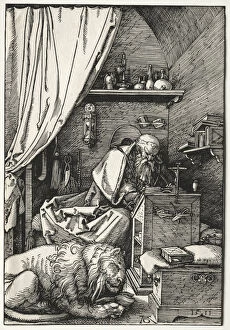
Background imageCell Collection: St. Jerome Cell 1511 Albrecht Dürer German

St. Jerome Cell 1511 Albrecht Dürer German
St. Jerome in His Cell, 1511. Albrecht Durer (German, 1471-1528). Woodcut

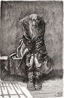
Background imageCell Collection: Fagin In The Condemned Cell. 'He Sat Down On A Stone Bench Opposite The Door

Fagin In The Condemned Cell. "He Sat Down On A Stone Bench Opposite The Door
Fagin In The Condemned Cell. " He Sat Down On A Stone Bench Opposite The Door, Which Served For A Seat And Bedstead, And Casting His Bloodshot Eyes Upon The Grond, Tried To Collect His Thoughts

Background imageCell Collection: Joan Of Arc 1412 To 1431 Being Interrogated In Her Cell By The Cardinal Of Winchester After The

Joan Of Arc 1412 To 1431 Being Interrogated In Her Cell By The Cardinal Of Winchester After The Painting By Paul Delaroche From The National And Domestic History Of England By William Aubrey

Background imageCell Collection: Sir Thomas More 1478 To 1535 In Prison. From The National And Domestic History Of England By

Sir Thomas More 1478 To 1535 In Prison. From The National And Domestic History Of England By William Aubrey Published London Circa 1890

Background imageCell Collection: Monotone Close Up Of Leaf

Monotone Close Up Of Leaf

Background imageCell Collection: A Man Stands In The Ocean With Items From Work And Vacation Flying Over His Head; Tarifa, Cadiz

A Man Stands In The Ocean With Items From Work And Vacation Flying Over His Head; Tarifa, Cadiz, Andalusia, Spain

Background imageCell Collection: Teenagers Studying The Bible

Teenagers Studying The Bible

Background imageCell Collection: Orbicular Design

Orbicular Design

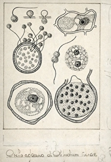
Background imageCell Collection: Botany, Fungi, Life cycle of Olpidium viciae, illustration

Botany, Fungi, Life cycle of Olpidium viciae, illustration
Botany - Fungi - Life cycle of Olpidium viciae. Illustration

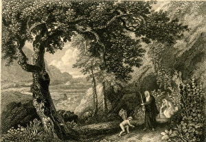
Background imageCell Collection: Shakespeare, The Tempest, Prosperos Cell

Shakespeare, The Tempest, Prosperos Cell on the enchanted island. With Prospero, Miranda and Ariel. 19th century

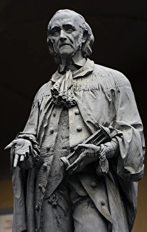
Background imageCell Collection: Alessandro Giuseppe Antonio Anastasio Volta (1745-1827). Ita

Alessandro Giuseppe Antonio Anastasio Volta (1745-1827). Italian physicist known for the invention of the battery in the 1800s. Statue. University of Pavia. Italy
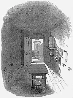
Background imageCell Collection: Parkhurst Prison, Isle of Wight - Cell

Parkhurst Prison, Isle of Wight - Cell
A cell at the juvenile penitentiary established in 1838 at Parkhurst, Isle of Wight. The prison aimed to provide boys with discipline, education and training prior to transportation. Date: circa 1847

Background imageCell Collection: Stone-breaking cell in a workhouse

Stone-breaking cell in a workhouse
Illustration showing a workhouse stone-breaking cell. Tramps and vagrants broke stones in return for a nights accommodation Date: circa 1909

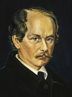
Background imageCell Collection: SCHLEIDEN, Matthias Jakob (1804-1881). German botanist

SCHLEIDEN, Matthias Jakob (1804-1881). German botanist and co-founder of the cell theory. Oil

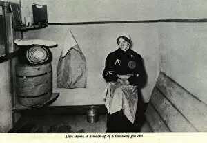
Background imageCell Collection: Elsie Howey Womens Exhibition Prison Cell

Elsie Howey Womens Exhibition Prison Cell. Elsie Howey (1884-1963) in a replica prison cell, dressed in mock prison uniform, at the Womens Exhibition, 13th-26th May 1909. Date: 1909

Background imageCell Collection: Suffragette Votes for Sheep Martyr

Suffragette Votes for Sheep Martyr. Little Bo Peep stands with her shepherds crook holding a picture of a sheep in a prison cell, with Votes for Women as the headline, and a martyr beneath

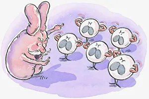
Background imageCell Collection: Cartoon representing thymus pointing at white blood cells flexing muscles

Cartoon representing thymus pointing at white blood cells flexing muscles

Background imageCell Collection: Conceptual image of platelet, red blood cell and white blood cell

Conceptual image of platelet, red blood cell and white blood cell

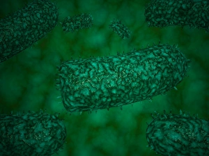
Background imageCell Collection: Stylized rabies virus particles

Stylized rabies virus particles, the cause of the viral neuroinvasive disease acute encephalitis

Background imageCell Collection: Microscopic view of yellow fever virus

Microscopic view of yellow fever virus. Yellow fever is an acute viral disease

Background imageCell Collection: Detailed cutaway diagram of human skin

Detailed cutaway diagram of human skin

Background imageCell Collection: Microscopic view of Giardiasis

Microscopic view of Giardiasis, an infectious disease caused by a unicellular parasite known as Giardia lamblia

Background imageCell Collection: Different types of acne, non-inflammatory and inflammatory

Different types of acne, non-inflammatory and inflammatory

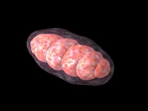
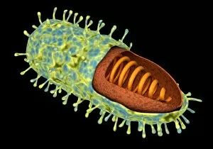
Background imageCell Collection: Conceptual image of mitochondria

Conceptual image of mitochondria

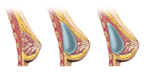
Background imageCell Collection: Woman breast implant cross section

Woman breast implant cross section. Comparison diagram, under and over pectoral muscle

Background imageCell Collection: Conceptual image of the layers of human skin

Conceptual image of the layers of human skin

Background imageCell Collection: Anatomy of foot fungus with microscopic close-up

Anatomy of foot fungus with microscopic close-up

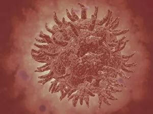
Background imageCell Collection: Conceptual image of rabies virus

Conceptual image of rabies virus

Background imageCell Collection: Madame de Guiche and her Daughter Appear at Night before the Artist in Prison, 1804

Madame de Guiche and her Daughter Appear at Night before the Artist in Prison, 1804
XIR173700 Madame de Guiche and her Daughter Appear at Night before the Artist in Prison, 1804 (w/c on paper) by Polignac, Armand de (1771-1847); 42.4x28 cm; Musee de la Ville de Paris

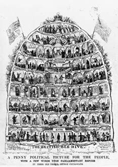
Background imageCell Collection: Peoples Beehive

Peoples Beehive
1840: The British Beehive, a cartoon depicting the population as members of a behive with the Queen at the top and various other occupations in cells below her

Background imageCell Collection: Police Station at Cherat - NWFP

Police Station at Cherat - NWFP. Cherat is a hill station, immediately above the village Chapri and Saleh Khana in the Nowshera District of Khyber-Pakhtunkhwa in Pakistan

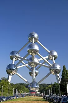
Background imageCell Collection: The Atomium monument at Brussels, Belgium

The Atomium monument at Brussels, Belgium

Background imageCell Collection: Picture No. 10876987

Picture No. 10876987
Scanning Electron Micrograph (SEM): Human skin section across vein showing red blood cells Date:

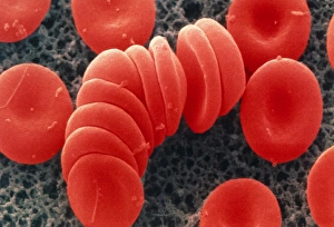
Background imageCell Collection: Coloured SEM of red blood cells, rouleau formation

Coloured SEM of red blood cells, rouleau formation
Red blood cells. Coloured scanning electron micrograph of red blood cells in rouleau formation. They often stack together in this way because of their shape

Background imageCell Collection: Paramecium sp. protozoan, SEM

Paramecium sp. protozoan, SEM
Paramecium protozoan. Coloured scanning electron micrograph (SEM) of a Paramecium sp. protozoan. This single-celled organism is aquatic, living in freshwater habitats

Background imageCell Collection: Euglena gracilis, SEM

Euglena gracilis, SEM
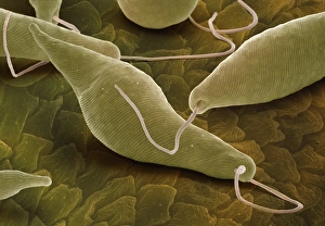
Euglena gracilis protists. Coloured scanning electron micrograph (SEM) of Euglena gracilis protists. These organisms possess a unique combination of plant and animal characters. Like the algae, E

Background imageCell Collection: Nasal lining, SEM

Nasal lining, SEM
Nasal lining. Coloured scanning electron micrograph (SEM) of the olfactory epithelium that lines the nasal cavity, showing olfactory cells (red) surrounded by numerous cilia (hair-like projections)

Background imageCell Collection: Zygote, artwork F006 / 2563

Zygote, artwork F006 / 2563
Zygote. Computer artwork of a fertilised ovum (egg), or zygote

Background imageCell Collection: Cell membrane lipid bilayer, artwork F007 / 1477

Cell membrane lipid bilayer, artwork F007 / 1477
Phospholipid bilayer. Computer artwork of the phospholipid bilayer that forms the membrane around all living cells. The cell membrane is made of phospholipid molecules

Background imageCell Collection: Osteocyte bone cell, SEM C016 / 9026

Osteocyte bone cell, SEM C016 / 9026
Osteocyte bone cell. Coloured scanning electron micrograph (SEM) of an osteocyte bone cell (red) surrounded by bone tissue (grey)

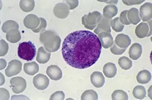
Background imageCell Collection: Myeloblast blood cell, light micrograph

Myeloblast blood cell, light micrograph
Myeloblast blood cell. Light micrograph of blood cells, including a myeloblast, a precursor for a type of white blood cell (leucocyte). Myeloblasts differentiate into granulocytes

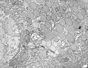
Background imageCell Collection: Pancreatic acinar cell

Pancreatic acinar cell. Transmission electron micrograph (TEM) of a section through an enzyme-secreting acinar cell in the human pancreas, showing part of the nucleus (round, far left)

Background imageCell Collection: Promyelocyte blood cell, light micrograph

Promyelocyte blood cell, light micrograph. This blood cell (centre) is a precursor for a type of white blood cell called a granulocyte, formed by granulopoiesis in the bone marrow

Background imageCell Collection: Chronic lymphocytic leukaemia, micrograph

Chronic lymphocytic leukaemia, micrograph
Chronic lymphocytic leukaemia. Light micrograph of blood cells sampled from a lymphatic ganglion in a case of chronic lymphocytic leukaemia, also called chronic lymphoid leukaemia (CLL)

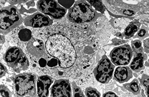
Background imageCell Collection: Macrophage and lymphocytes, TEM

Macrophage and lymphocytes, TEM
Macrophage and lymphocytes, transmission electron micrograph (TEM). Macrophage surrounded by lymphocytes in a lymph node. This view includes many intercellular contacts

Background imageCell Collection: Hepatocyte liver cell, TEM

Hepatocyte liver cell, TEM
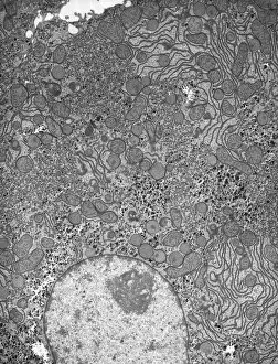
Hepatocyte liver cell. Transmission electron micrograph (TEM) of a section through an hepatocyte liver cell, showing rough and smooth endoplasmic reticulum (ER, dark lines)

Background imageCell Collection: Liver tissue, TEM

Liver tissue, TEM
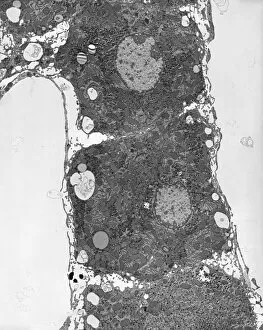
Liver tissue. Transmission electron micrograph (TEM) of a section through the liver, showing part of a radial cord of hepatocyte liver cells (dark) and the vascular sinusoids (white)


All Professionally Made to Order for Quick Shipping

"Exploring the Intricacies of Life: Unveiling the Wonders of Cells" Delving into the microscopic world, a histological diagram of a mammalian retina reveals the intricate structure and organization of cells that enable us to perceive light. The cerebellum tissue, captured in a light micrograph, showcases its complex network of cells responsible for coordinating movement and balance. Intriguingly, even philosophers find solace in contemplating cellular existence. "Philosopher in Meditation, " an oil painting from 1632, reminds us that pondering life's mysteries often leads us back to our fundamental building blocks – cells. Zooming further into this realm, nerve and glial cells come alive under the lens. This captivating light micrograph highlights their vital role in transmitting signals throughout our nervous system. Meanwhile, a lavender pollen grain captured through scanning electron microscopy (SEM) displays nature's exquisite design at the cellular level. The battle against diseases takes center stage as T lymphocytes confront cancer cells in another SEM image. Science and evolution intertwine as we witness these tiny warriors fighting for survival within our bodies. Artistic expressions also shed light on cell significance; "The Death of Socrates, " painted in 1787, serves as a poignant reminder that even great minds are ultimately composed of countless individual cells. Santiago Ramon y Cajal's meticulous drawing from 1894 unveils various cell types within the mammalian cerebellum. His work not only represents scientific progress but also exemplifies how art can aid understanding by visually capturing complexity. Stepping away from biology momentarily, we encounter Rolls Royce/Snecma Olympus 593 Mk602 engine undergoing testing within a controlled environment called a test cell. Here too, precision engineering relies on understanding cellular mechanics to achieve optimal performance. Returning to biological marvels - HeLa cells take center stage under yet another microscope lens. These immortalized human cells have revolutionized medical research, paving the way for countless breakthroughs.