Fraud Blocker Skip to main content

Cell Collection (page 3)

"Exploring the Intricacies of Life: Unveiling the Wonders of Cells" Delving into the microscopic world

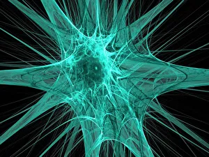
Background imageCell Collection: Nerve cells, abstract artwork

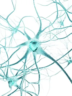
Nerve cells, abstract artwork
Nerve cells. Abstract computer artwork of nerve cells, or neurons. Neurons are responsible for passing information around the central nervous system (CNS) and from the CNS to the rest of the body
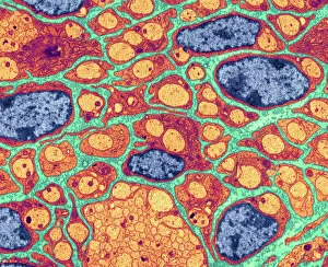
Background imageCell Collection: Myelination of nerve fibres, TEM

Myelination of nerve fibres, TEM
Myelination of nerve fibres. Coloured transmission electron micrograph (TEM) of Schwann cells (red, with blue nuclei) insulating nerve fibres (axons, orange) with a myelin sheath

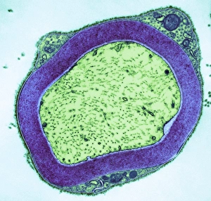
Background imageCell Collection: Myelinated nerve, TEM

Myelinated nerve, TEM
Myelinated nerve. Coloured transmission electron micrograph (TEM) of myelinated nerve fibres and Schwann cells. Myelin (purple) is an insulating fatty layer that surrounds nerve fibres (axons)

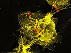
Background imageCell Collection: Neutrophil cell trapping bacteria, SEM

Neutrophil cell trapping bacteria, SEM
Neutrophil cell trapping bacteria. Coloured scanning electron micrograph (SEM) of bacteria (rod-shaped) being trapped by a neutrophil cell

Background imageCell Collection: Red blood cells and heart

Red blood cells and heart. Computer artwork of a heart on the silhouette of a person and red blood cells (erythrocytes). The heart is a hollow muscle that pumps blood around the body

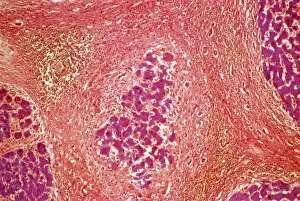
Background imageCell Collection: Liver tissue cirrhosis, light micrograph

Liver tissue cirrhosis, light micrograph
Liver cirrhosis. Light micrograph of a section through liver tissue showing cirrhosis. This is a condition where the liver responds to the injury or death of some of its cells by producing

Background imageCell Collection: Mitochondrion, TEM

Mitochondrion, TEM
Mitochondrion. Coloured transmission electron micrograph (TEM) of mitochondria (green/blue) in heart muscle. Mitochondria are organelles found in the cytoplasm of eukaryotic cells

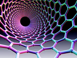
Background imageCell Collection: Carbon nanotube

Carbon nanotube. Computer artwork showing the hexagonal carbon structure of a nanotube, or buckytube

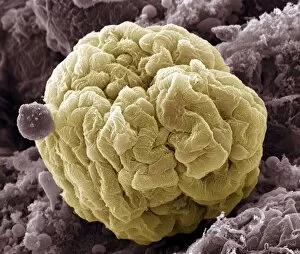
Background imageCell Collection: Kidney glomerulus, SEM

Kidney glomerulus, SEM
Kidney glomerulus, coloured scanning electron micrograph (SEM). Podocyte cells, which make up the surface of the glomerulus, are yellow. Magnification: x650 when printed at 10 centimetres wide

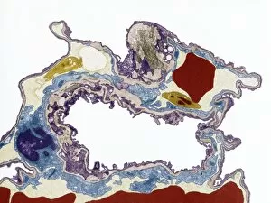
Background imageCell Collection: Lung tissue, TEM

Lung tissue, TEM
Lung tissue, coloured transmission electron micrograph (TEM). A single alveolus is at center. At top right is a pulmonary capillary containing a red blood cell (red) and a platelet (yellow)

Background imageCell Collection: Fibroblast cell, artwork

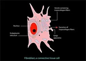
Fibroblast cell, artwork
Fibroblast cell. Computer artwork of a fibroblast excreting collagen fibres (tropocollagen). Fibroblasts are cells that produce connective tissue such as collagen (tropocollagen)

Background imageCell Collection: Trachea lining, SEM

Trachea lining, SEM
Trachea lining, coloured scanning electron micrograph (SEM). The tracheal lining contains mucus-secreting goblet cells and epithelial cells bearing hair-like cilia (blue)

Background imageCell Collection: Cotton plant leaf, light micrograph

Cotton plant leaf, light micrograph
Cotton plant leaf. Light micrograph of a transverse section through a leaf of the cotton plant (Gossypium hirsutum). Magnification: x45 when printed at 10 centimetres wide

Background imageCell Collection: Silver birch twig, SEM

Silver birch twig, SEM
Silver birch twig. Coloured scanning electron micrograph (SEM) of a section through a silver birch (Betula pendula) twig. At the centre of the twig (top centre left) is the pith

Background imageCell Collection: Pseudomonas aeruginosa bacteria, SEM

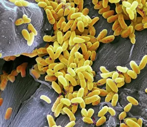
Pseudomonas aeruginosa bacteria, SEM
Pseudomonas aeruginosa bacteria, coloured scanning electron micrograph (SEM). These Gram-negative rod-shaped bacteria are found in soil and water, and as normal flora in the human intestine

Background imageCell Collection: Fagin in Jail

Fagin in Jail
Fagin in the condemned cell

Background imageCell Collection: Fagin in the condemned cell (w / c on paper)

Fagin in the condemned cell (w / c on paper)
614693 Fagin in the condemned cell (w/c on paper) by English School, (19th century); Private Collection; (add.info.: Fagin in the condemned cell)

Background imageCell Collection: St Jerome in His Study (engraving)

St Jerome in His Study (engraving)
3646436 St Jerome in His Study (engraving) by Durer or Duerer, Albrecht (1471-1528) (after); Private Collection; (add.info.: St Jerome in His Study)

Background imageCell Collection: Osteocyte bone cell, SEM

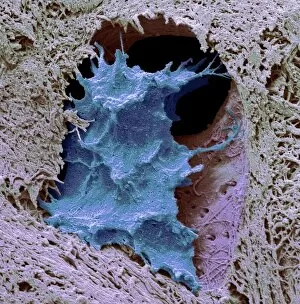
Osteocyte bone cell, SEM
Osteocyte bone cell. Coloured scanning electron micrograph (SEM) of an osteocyte bone cell (blue) surrounded by bone tissue (grey)

Background imageCell Collection: Suffragette in Prison Cell

Suffragette in Prison Cell. A suffragette languishes in a bare prison cell. Captioned, Edie - A Good Suff! The drawing is initialed E.S.P - E. Sylvia Pankhurst? Date: circa 1909

Background imageCell Collection: Nerve cell, artwork F007 / 7448

Nerve cell, artwork F007 / 7448
Nerve cell, computer artwork

Background imageCell Collection: Sleeping cell at Brixton Prison

Sleeping cell at Brixton Prison

Background imageCell Collection: Dinoflagellates, SEM

Dinoflagellates, SEM
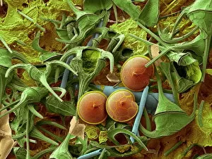
Dinoflagellates. Coloured scanning electron micrograph (SEM) of Prorocentrum dinoflagellates (round) and Ceratium dinoflagellates (branched). Dinoflagellates are unicellular protozoans

Background imageCell Collection: Red blood cells, SEM

Red blood cells, SEM
Red blood cells. Coloured scanning electron micrograph (SEM) of red blood cells (erythrocytes). Red blood cells are biconcave, disc-shaped cells that transport oxygen from the lungs to body cells

Background imageCell Collection: Water fern rhizome, light micrograph

Water fern rhizome, light micrograph
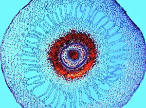
Water fern rhizome. Polarised light micrograph of a section through a rhizome from a water fern (Marselia vestita). Under the epidermis (the outer layer)

Background imageCell Collection: Diatom cell wall, SEM

Diatom cell wall, SEM
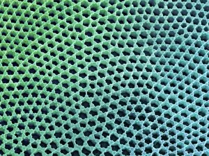
Diatom cell wall. Coloured scanning electron micrograph (SEM) of the pattern of small holes, called striae, in the cell wall of a diatom alga. This is a Coscinodiscus sp. diatom

Background imageCell Collection: Corridor and cell doors, Bodmin Gaol, Cornwall

Corridor and cell doors, Bodmin Gaol, Cornwall
The main corridor and cell doors at Bodmin Gaol, Cornwall

Background imageCell Collection: Blastocyst, artwork F006 / 2190

Blastocyst, artwork F006 / 2190
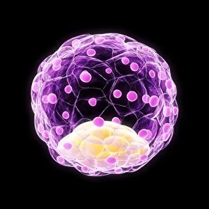
Blastocyst. Computer artwork of a 100-cell blastocyst embryo

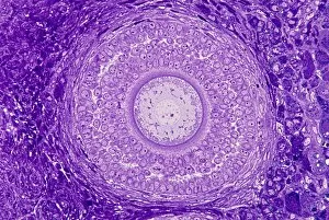
Background imageCell Collection: Ovarian follicle, light micrograph C016 / 0519

Ovarian follicle, light micrograph C016 / 0519
Ovarian follicle. Light micrograph of a section through a secondary ovarian follicle showing signs of maturation into the antral follicle class

Background imageCell Collection: Eye muscle, TEM C014 / 1468

Eye muscle, TEM C014 / 1468
Eye muscle. Transmission electron micrograph (TEM) of a section through a striated muscle cell from the ciliary muscle of a human eye

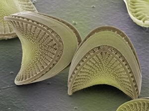
Background imageCell Collection: Diatoms, SEM

Diatoms, SEM
Diatoms. Coloured scanning electron micrograph (SEM) of diatoms (Campylodiscus sp.). These microscopic, single-celled algae possess rigid and finely-sculpted cell walls known as frustules

Background imageCell Collection: Coloured SEM of a leukaemic white blood cell M132 / 0488

Coloured SEM of a leukaemic white blood cell M132 / 0488
Leukaemia cell. Coloured scanning electron micrograph (SEM) of a cancerous white blood cell in acute myeloid leukaemia. Leukaemia is a cancer of the blood which results in the over-production of

Background imageCell Collection: Foraminifer model

Foraminifer model
Model of typical nummulitic foraminfer after Zittel

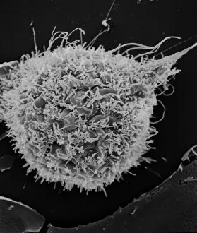
Background imageCell Collection: T2 cell culture

T2 cell culture
Scanning electron microscope image showing a T2 cell culture (x 4K)

Background imageCell Collection: Skin layers, SEM

Skin layers, SEM

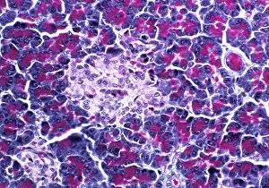
Background imageCell Collection: Pancreatic islet of Langerhans

Pancreatic islet of Langerhans
Islet of Langerhans. Light micrograph of a section through an islet of Langerhans (pale, centre) in pancreas tissue. This clump of secretory cells forms part of the endocrine system of the body

Background imageCell Collection: Astrocyte nerve cell

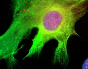
Astrocyte nerve cell. Fluorescent light micrograph of an astrocyte cell from a human brain. Intermediate filaments (IFs), part of the cells cytoskeleton, have been dyed green

Background imageCell Collection: Blood clot on plaster, SEM

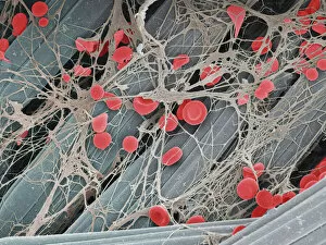
Blood clot on plaster, SEM
Blood clot on plaster. Coloured scanning electron micrograph (SEM) of blood clotting on the surface of a sticking plaster used to dress a small cut

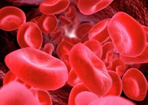
Background imageCell Collection: Red blood cells, computer artwork

Red blood cells, computer artwork
Red blood cells. Computer artwork of human red blood cells (erythrocytes) in a blood vessel. Red blood cells are biconcave, giving them a large surface area for gas exchange, and highly elastic

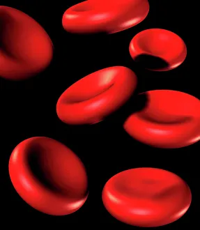
Background imageCell Collection: Red blood cells

Red blood cells. Computer artwork of human red blood cells (erythrocytes). Red blood cells are biconcave, giving them a large surface area for gas exchange, and highly elastic

Background imageCell Collection: Rift Valley fever virus, TEM

Rift Valley fever virus, TEM

Background imageCell Collection: Vesicular stomatitis virus, TEM

Vesicular stomatitis virus, TEM
Vesicular stomatitis virus. Coloured transmission electron micrograph (TEM) of vesicular stomatitis virus (VSV) particles (blue) budding from a host cell (red)

Background imageCell Collection: Stem cell, SEM

Stem cell, SEM
Stem cell, coloured scanning electron micrograph (SEM). Stem cells can differentiate into any other cell type. There are three main types of mammalian stem cell: embryonic stem cells

Background imageCell Collection: Stem cells, SEM

Stem cells, SEM
Stem cells, coloured scanning electron micrograph (SEM). Stem cells can differentiate into any other cell type. There are three main types of mammalian stem cell: embryonic stem cells

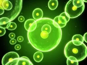
Background imageCell Collection: Cells, conceptual computer artwork

Cells, conceptual computer artwork

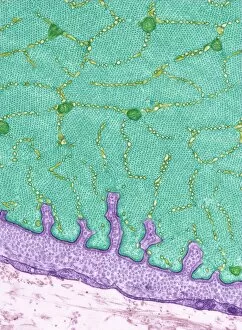
Background imageCell Collection: Intestinal microvilli, TEM

Intestinal microvilli, TEM
Intestinal microvilli, coloured transmission electron micrograph (TEM). The microvilli are finger-like projections (seen here in circular cross-section)

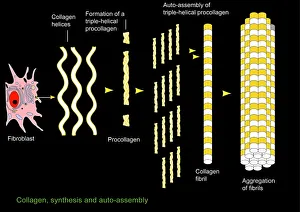
Background imageCell Collection: Collagen synthesis and assembly, artwork

Collagen synthesis and assembly, artwork. At left is a fibroblast, the cell that synthesises helical protein chains of collagen (wavy lines)


All Professionally Made to Order for Quick Shipping

"Exploring the Intricacies of Life: Unveiling the Wonders of Cells" Delving into the microscopic world, a histological diagram of a mammalian retina reveals the intricate structure and organization of cells that enable us to perceive light. The cerebellum tissue, captured in a light micrograph, showcases its complex network of cells responsible for coordinating movement and balance. Intriguingly, even philosophers find solace in contemplating cellular existence. "Philosopher in Meditation, " an oil painting from 1632, reminds us that pondering life's mysteries often leads us back to our fundamental building blocks – cells. Zooming further into this realm, nerve and glial cells come alive under the lens. This captivating light micrograph highlights their vital role in transmitting signals throughout our nervous system. Meanwhile, a lavender pollen grain captured through scanning electron microscopy (SEM) displays nature's exquisite design at the cellular level. The battle against diseases takes center stage as T lymphocytes confront cancer cells in another SEM image. Science and evolution intertwine as we witness these tiny warriors fighting for survival within our bodies. Artistic expressions also shed light on cell significance; "The Death of Socrates, " painted in 1787, serves as a poignant reminder that even great minds are ultimately composed of countless individual cells. Santiago Ramon y Cajal's meticulous drawing from 1894 unveils various cell types within the mammalian cerebellum. His work not only represents scientific progress but also exemplifies how art can aid understanding by visually capturing complexity. Stepping away from biology momentarily, we encounter Rolls Royce/Snecma Olympus 593 Mk602 engine undergoing testing within a controlled environment called a test cell. Here too, precision engineering relies on understanding cellular mechanics to achieve optimal performance. Returning to biological marvels - HeLa cells take center stage under yet another microscope lens. These immortalized human cells have revolutionized medical research, paving the way for countless breakthroughs.