Fraud Blocker Skip to main content

Cell Collection (page 4)

"Exploring the Intricacies of Life: Unveiling the Wonders of Cells" Delving into the microscopic world

Background imageCell Collection: Stem cells, SEM

Stem cells, SEM
Stem cells, coloured scanning electron micrograph (SEM). Stem cells can differentiate into any other cell type. There are three main types of mammalian stem cell: embryonic stem cells

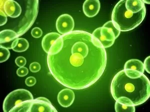
Background imageCell Collection: Cells, conceptual computer artwork

Cells, conceptual computer artwork

Background imageCell Collection: Intestinal microvilli, TEM

Intestinal microvilli, TEM
Intestinal microvilli, coloured transmission electron micrograph (TEM). The microvilli are finger-like projections (seen here in circular cross-section)

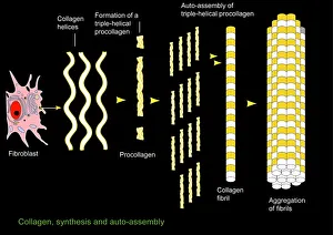
Background imageCell Collection: Collagen synthesis and assembly, artwork

Collagen synthesis and assembly, artwork. At left is a fibroblast, the cell that synthesises helical protein chains of collagen (wavy lines)

Background imageCell Collection: Skin surface, SEM

Skin surface, SEM
Skin surface. Coloured scanning electron micrograph (SEM) of squamous cells on the surface of the skin. These are flat, keratinised

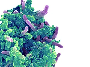
Background imageCell Collection: Bacteria infecting a macrophage, SEM

Bacteria infecting a macrophage, SEM
Bacteria infecting a macrophage. Coloured scanning electron micrograph (SEM) of Mycobacterium tuberculosis bacteria (purple) infecting a macrophage white blood cell

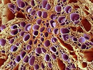
Background imageCell Collection: Xylem tissue, SEM

Xylem tissue, SEM
Xylem tissue. Coloured scanning electron micrograph (SEM) of a section through xylem tissue from a dicotyledon rootlet. Xylem vessels (purple)

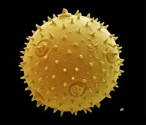
Background imageCell Collection: Marrow and forget-me-not pollen, SEM

Marrow and forget-me-not pollen, SEM
Pollen grains. Coloured scanning electron micrograph (SEM) of a pollen grain from a marrow plant (Cucurbita sp. left) and a forget-me-not plant (Myosotis sp. lower right)
Background imageCell Collection: Wood, light micrograph

Wood, light micrograph
Wood. Light micrograph of a transverse section through wood showing annual growth rings (near horizontal bands). A years growth consists of a porous band, which is the slow winter growth

Background imageCell Collection: Stonehenge / Stukeley

Stonehenge / Stukeley
A peep into the Sanctum Sanctorum. Stukeley states When we advance.. the chasm of sky between the jambs of the cell.. and the greatness of every part, surprizes

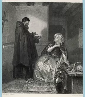
Background imageCell Collection: Romeo & Juliet / Friar

Romeo & Juliet / Friar
Act IV Juliet visits Friar Lawrence in his cell and procures a sleeping potion

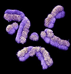
Background imageCell Collection: Human chromosomes, SEM C013 / 5002

Human chromosomes, SEM C013 / 5002
Human chromosomes. Coloured scanning electron micrograph (SEM) of human chromosomes prepared with the harlequin staining technique

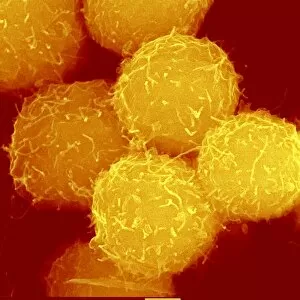
Background imageCell Collection: Haematopoietic stem cells, SEM C013 / 5009

Haematopoietic stem cells, SEM C013 / 5009
Haematopoietic stem cells, coloured scanning electron micrograph (SEM). Stem cells can differentiate into any other cell type

Background imageCell Collection: Show You My Selfie

Show You My Selfie
Dikhotomy

Background imageCell Collection: Haunting Thoughts

Haunting Thoughts
Petri Damstén

Background imageCell Collection: Guillotine

Guillotine
Petri Damstén

Background imageCell Collection: Handle with Care

Handle with Care
Petri Damstén

Background imageCell Collection: Kiss of Death

Kiss of Death
Petri Damstén

Background imageCell Collection: Waiting for the Coffin

Waiting for the Coffin
Petri Damstén

Background imageCell Collection: Eternal Pain

Eternal Pain
Petri Damstén

Background imageCell Collection: Backstab

Backstab
Petri Damstén

Background imageCell Collection: The Endless Rain

The Endless Rain
Petri Damstén

Background imageCell Collection: Roles I Have to Play

Roles I Have to Play
Petri Damstén

Background imageCell Collection: Trust You Seek

Trust You Seek
Petri Damstén

Background imageCell Collection: Show You My Selfie

Show You My Selfie
Dikhotomy

Background imageCell Collection: Show You My Selfie

Show You My Selfie
Dikhotomy

Background imageCell Collection: Punishment Cell, 1891. Creator: Aleksei Kuznetsov

Punishment Cell, 1891. Creator: Aleksei Kuznetsov
Punishment Cell, 1891. One of 74 views taken in July 1891 and contained in the albumTipy i vidy Nerchinskoi katorgi (Views and inhabitants of Nerchinsk hard labor camps)

Background imageCell Collection: Pregnant nun Sister Jean sitting in her convent cell

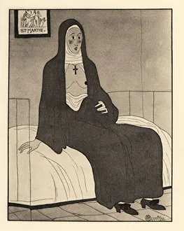
Pregnant nun Sister Jean sitting in her convent cell, her habit open to reveal her breasts. A picture of Saint Martha on the wall. Soeur Jeanne

Background imageCell Collection: Yeast cells, SEM

Yeast cells, SEM
Yeast cells. Coloured scanning electron micrograph (SEM) of cells of brewer s, or baker s, yeast (Saccharomyces cerevisiae). This fungus consists of single vegetative cells

Background imageCell Collection: SCHLEIDEN, Matthias Jakob (1804-1881). German

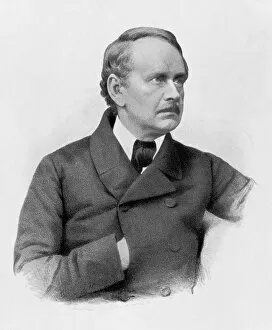
SCHLEIDEN, Matthias Jakob (1804-1881). German botanist and co-founder of the cell theory. Litography

Background imageCell Collection: Specimen XII

Specimen XII
Amy Louise Baker

Background imageCell Collection: Specimen XI

Specimen XI
Amy Louise Baker

Background imageCell Collection: Specimen X

Specimen X
Amy Louise Baker

Background imageCell Collection: Specimen IX

Specimen IX
Amy Louise Baker

Background imageCell Collection: Specimen VIII

Specimen VIII
Amy Louise Baker

Background imageCell Collection: Specimen VII

Specimen VII
Amy Louise Baker

Background imageCell Collection: Specimen VI

Specimen VI
Amy Louise Baker

Background imageCell Collection: Specimen V

Specimen V
Amy Louise Baker

Background imageCell Collection: Specimen IV

Specimen IV
Amy Louise Baker

Background imageCell Collection: Specimen III

Specimen III
Amy Louise Baker

Background imageCell Collection: Specimen II

Specimen II
Amy Louise Baker

Background imageCell Collection: Specimen I

Specimen I
Amy Louise Baker

Background imageCell Collection: Meryemana Manastiri: the monastery in Sumela, Trebisonda Territory, Turkey

Meryemana Manastiri: the monastery in Sumela, Trebisonda Territory, Turkey Turkey Sumela. Date of Photograph:1976-1977

Background imageCell Collection: Study for the 'Liberation of St. Peter', drawing, Gabinetto dei Disegni e delle Stampe

Study for the "Liberation of St. Peter", drawing, Gabinetto dei Disegni e delle Stampe, Uffizi Gallery, Florence Florence Uffizi Gallery

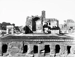
Background imageCell Collection: General view of the remains of the Temple of Venus and Rome, Hadrian work preserved in Rome

General view of the remains of the Temple of Venus and Rome, Hadrian work preserved in Rome Rome Temple Hadrianic Period, Imperial Period, Roman Art, Europe, Ancient Civilization

Background imageCell Collection: The Temple of Venus and Rome, Hadrian work preserved in Rome

The Temple of Venus and Rome, Hadrian work preserved in Rome Rome Temple Hadrianic Period, Imperial Period, Roman Art, Europe, Ancient Civilization. Date of Photograph:1880-1890 ca

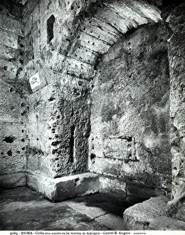
Background imageCell Collection: Detail of the cell where the tomb of the Emperor Hadrian was located in the Castle of S

Detail of the cell where the tomb of the Emperor Hadrian was located in the Castle of S. Angelo, Rome. Rome Castle of S. Angelo Castle, Mausoleum Hadrianic Period, Imperial Period, Roman Art, Europe

Background imageCell Collection: Mamertino Prison (called Tullianum in ancient times), today the Chapel of San Pietro in Carcere

Mamertino Prison (called Tullianum in ancient times), today the Chapel of San Pietro in Carcere, located under the Church of S. Giuseppe dei Falegnami in Rome


All Professionally Made to Order for Quick Shipping

"Exploring the Intricacies of Life: Unveiling the Wonders of Cells" Delving into the microscopic world, a histological diagram of a mammalian retina reveals the intricate structure and organization of cells that enable us to perceive light. The cerebellum tissue, captured in a light micrograph, showcases its complex network of cells responsible for coordinating movement and balance. Intriguingly, even philosophers find solace in contemplating cellular existence. "Philosopher in Meditation, " an oil painting from 1632, reminds us that pondering life's mysteries often leads us back to our fundamental building blocks – cells. Zooming further into this realm, nerve and glial cells come alive under the lens. This captivating light micrograph highlights their vital role in transmitting signals throughout our nervous system. Meanwhile, a lavender pollen grain captured through scanning electron microscopy (SEM) displays nature's exquisite design at the cellular level. The battle against diseases takes center stage as T lymphocytes confront cancer cells in another SEM image. Science and evolution intertwine as we witness these tiny warriors fighting for survival within our bodies. Artistic expressions also shed light on cell significance; "The Death of Socrates, " painted in 1787, serves as a poignant reminder that even great minds are ultimately composed of countless individual cells. Santiago Ramon y Cajal's meticulous drawing from 1894 unveils various cell types within the mammalian cerebellum. His work not only represents scientific progress but also exemplifies how art can aid understanding by visually capturing complexity. Stepping away from biology momentarily, we encounter Rolls Royce/Snecma Olympus 593 Mk602 engine undergoing testing within a controlled environment called a test cell. Here too, precision engineering relies on understanding cellular mechanics to achieve optimal performance. Returning to biological marvels - HeLa cells take center stage under yet another microscope lens. These immortalized human cells have revolutionized medical research, paving the way for countless breakthroughs.