Fraud Blocker Skip to main content

Cellular Collection

"Unveiling the Intricacies of Cellular Life: A Journey Through Micrographs" Budding yeast cell: Witness the birth of new life as a budding yeast cell emerges

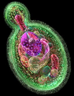
Background imageCellular Collection: Budding yeast cell

Budding yeast cell. Computer artwork of asection through a yeast ( Candida albicans )cell that is reproducing asexually. A daughtercell (top left) is budding from the parent cell

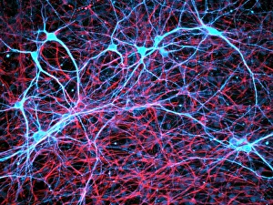
Background imageCellular Collection: Nerve and glial cells, light micrograph

Nerve and glial cells, light micrograph
Nerve and glial cells, fluorescence light micrograph. These are neural stem cells that have differentiated into neurons (nerve cells, blue) and glial cells (support cells, red)

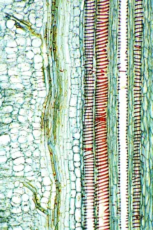
Background imageCellular Collection: Dicotyledon plant stem, light micrograph

Dicotyledon plant stem, light micrograph
Dicotyledon plant stem. Light micrograph of a longitudinal radial section through a typical dicotyledon stem. The section has passed through a vascular bundle and other stem tissue

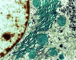
Background imageCellular Collection: Rough endoplasmic reticulum, TEM

Rough endoplasmic reticulum, TEM
Rough endoplasmic reticulum, coloured transmission electron micrograph (TEM). This section shows the rough endoplasmic reticulum (ER, folds, centre), a membranous structure that occurs in cells

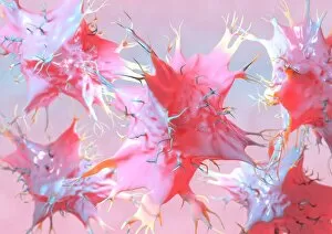
Background imageCellular Collection: Dendritic cells, artwork

Dendritic cells, artwork. Dendritic cells, a type of white blood cell, are called antigen presenting cells (APCs) and are found in the bodys tissues

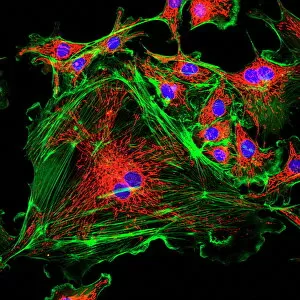
Background imageCellular Collection: Cell structure

Cell structure. Confocal light micrograph of cultured endothelial cells. A fluorescent dye has been used to show the cell structure
Background imageCellular Collection: Mitosis, light micrograph

Mitosis, light micrograph
Mitosis. Confocal light micrograph of the stages of mitosis (nuclear division) and cytokinesis (cell division). During mitosis the nuclear envelope disintegrates (3rd image) and the chromosomes (blue)

Background imageCellular Collection: Cell division, fluorescent micrograph

Cell division, fluorescent micrograph
Cell division. Immunofluorescent light micrograph of a human epithelial cell (centre) during the late anaphase stage of mitosis

Background imageCellular Collection: Dohle bodies in blood cell, micrograph

Dohle bodies in blood cell, micrograph
Dohle bodies in blood cell. Light micrograph of a neutrophil white blood cell (centre) with Dohle bodies. These are small inclusions within the cells cytoplasm

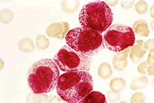
Background imageCellular Collection: Acute promyelocytic leukaemia, micrograph

Acute promyelocytic leukaemia, micrograph
Acute promyelocytic leukaemia. Light micrograph of blood cells from bone marrow in a case of acute promyelocytic leukaemia. Leukaemia is a cancer where certain blood cells form in excess

Background imageCellular Collection: Phagocytosis of fungal spores, SEM

Phagocytosis of fungal spores, SEM
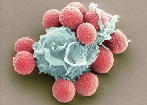
Phagocytosis of a fungus spore. Coloured scanning electron micrograph (SEM) of a neutrophil (blue) phagocytosing (engulfing and destroying) spores from the fungus Aspergillus fumigatus (red)

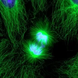
Background imageCellular Collection: Cell division, fluorescent micrograph

Cell division, fluorescent micrograph
Cell division. Immunofluorescent light micrograph of a human epithelial cell (centre) during the interphase stage of mitosis

Background imageCellular Collection: Trachea lining, SEM

Trachea lining, SEM
Trachea lining, coloured scanning electron micrograph (SEM). The tracheal lining contains mucus-secreting goblet cells and epithelial cells bearing hair-like cilia (blue)

Background imageCellular Collection: Cotton plant leaf, light micrograph

Cotton plant leaf, light micrograph
Cotton plant leaf. Light micrograph of a transverse section through a leaf of the cotton plant (Gossypium hirsutum). Magnification: x45 when printed at 10 centimetres wide

Background imageCellular Collection: Water fern rhizome, light micrograph

Water fern rhizome, light micrograph
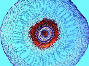
Water fern rhizome. Polarised light micrograph of a section through a rhizome from a water fern (Marselia vestita). Under the epidermis (the outer layer)

Background imageCellular Collection: Diatoms, SEM

Diatoms, SEM
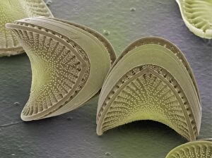
Diatoms. Coloured scanning electron micrograph (SEM) of diatoms (Campylodiscus sp.). These microscopic, single-celled algae possess rigid and finely-sculpted cell walls known as frustules

Background imageCellular Collection: Human chromosomes, SEM C013 / 5002

Human chromosomes, SEM C013 / 5002
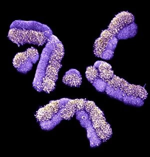
Human chromosomes. Coloured scanning electron micrograph (SEM) of human chromosomes prepared with the harlequin staining technique

Background imageCellular Collection: Haematopoietic stem cells, SEM C013 / 5009

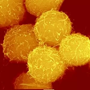
Haematopoietic stem cells, SEM C013 / 5009
Haematopoietic stem cells, coloured scanning electron micrograph (SEM). Stem cells can differentiate into any other cell type

Background imageCellular Collection: Colombia, Cartagena, Girl Taking Photo of Pegasus Sculpture

Colombia, Cartagena, Girl Taking Photo of Pegasus Sculpture

Background imageCellular Collection: Specimen XII

Specimen XII
Amy Louise Baker

Background imageCellular Collection: Specimen XI

Specimen XI
Amy Louise Baker

Background imageCellular Collection: Specimen X

Specimen X
Amy Louise Baker

Background imageCellular Collection: Specimen IX

Specimen IX
Amy Louise Baker

Background imageCellular Collection: Specimen VIII

Specimen VIII
Amy Louise Baker

Background imageCellular Collection: Specimen VII

Specimen VII
Amy Louise Baker

Background imageCellular Collection: Specimen VI

Specimen VI
Amy Louise Baker

Background imageCellular Collection: Specimen V

Specimen V
Amy Louise Baker

Background imageCellular Collection: Specimen IV

Specimen IV
Amy Louise Baker

Background imageCellular Collection: Specimen III

Specimen III
Amy Louise Baker

Background imageCellular Collection: Specimen II

Specimen II
Amy Louise Baker

Background imageCellular Collection: Specimen I

Specimen I
Amy Louise Baker

Background imageCellular Collection: Telecommunications Cell Phone Tower With Antenna In Countryside. Belarus. Aerial View

Telecommunications Cell Phone Tower With Antenna In Countryside. Belarus. Aerial View Telecommunications Cell Phone Tower With Antenna In Countryside. Belarus. Aerial View

Background imageCellular Collection: South East Asia, Thailand, Buddhist monk using a lap top and a mobile phone

South East Asia, Thailand, Buddhist monk using a lap top and a mobile phone

Background imageCellular Collection: Colony of Haemophilus influenzae, 1906 (litho)

Colony of Haemophilus influenzae, 1906 (litho)
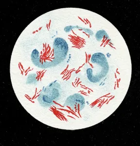
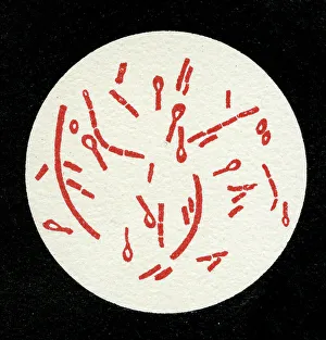
3501914 Colony of Haemophilus influenzae, 1906 (litho) by German School, (20th century); Private Collection; (add.info.: Bacteria constitute a large domain of prokaryotic microorganisms)

Background imageCellular Collection: Colony of Mycobacterium leprae, 1906 (litho)

Colony of Mycobacterium leprae, 1906 (litho)
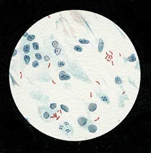
3501912 Colony of Mycobacterium leprae, 1906 (litho) by German School, (20th century); Private Collection; (add.info.: Bacteria constitute a large domain of prokaryotic microorganisms)

Background imageCellular Collection: Colony of Micrococcus Gonorrhoea, 1906 (litho)

Colony of Micrococcus Gonorrhoea, 1906 (litho)
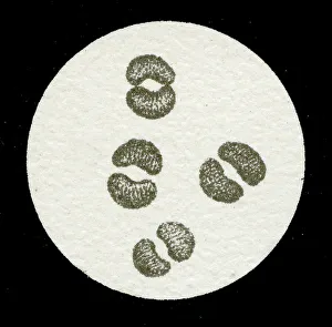
3501903 Colony of Micrococcus Gonorrhoea, 1906 (litho) by German School, (20th century); Private Collection; (add.info.: Bacteria constitute a large domain of prokaryotic microorganisms)

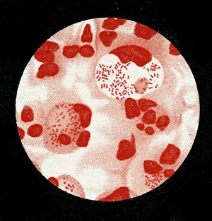
Background imageCellular Collection: Colony of Streptococcus pneumoniae with and without bubble capsule, 1906 (litho)

Colony of Streptococcus pneumoniae with and without bubble capsule, 1906 (litho)
3501899 Colony of Streptococcus pneumoniae with and without bubble capsule, 1906 (litho) by German School, (20th century); Private Collection; (add.info)

Background imageCellular Collection: Colony of Streptococcus Pneumoniae, 1906 (litho)

Colony of Streptococcus Pneumoniae, 1906 (litho)
3501898 Colony of Streptococcus Pneumoniae, 1906 (litho) by German School, (20th century); Private Collection; (add.info.: Bacteria constitute a large domain of prokaryotic microorganisms)

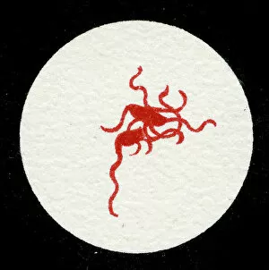
Background imageCellular Collection: Spirillum of the Lyme disease causing bacteria, Spirochaetes Borrelia Recurrentis

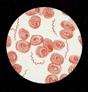
Spirillum of the Lyme disease causing bacteria, Spirochaetes Borrelia Recurrentis, in blood sample, 1906 (litho)
3501919 Spirillum of the Lyme disease causing bacteria, Spirochaetes Borrelia Recurrentis, in blood sample, 1906 (litho) by German School

Background imageCellular Collection: Colony of Vibrio cholerae, 1906 (litho)

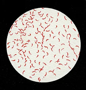
Colony of Vibrio cholerae, 1906 (litho)
3501918 Colony of Vibrio cholerae, 1906 (litho) by German School, (20th century); Private Collection; (add.info.: Bacteria constitute a large domain of prokaryotic microorganisms)

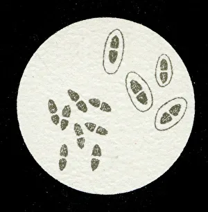
Background imageCellular Collection: Colony of Clostridium tetani with spores, 1906 (litho)

Colony of Clostridium tetani with spores, 1906 (litho)
3501913 Colony of Clostridium tetani with spores, 1906 (litho) by German School, (20th century); Private Collection; (add.info.: Bacteria constitute a large domain of prokaryotic microorganisms)

Background imageCellular Collection: Colony of Mycobacterium tuberculosis, 1906 (litho)

Colony of Mycobacterium tuberculosis, 1906 (litho)
3501910 Colony of Mycobacterium tuberculosis, 1906 (litho) by German School, (20th century); Private Collection; (add.info.: Bacteria constitute a large domain of prokaryotic microorganisms)

Background imageCellular Collection: A colony of Staphylococcus pyogenes after cell division, 1906 (litho)

A colony of Staphylococcus pyogenes after cell division, 1906 (litho)
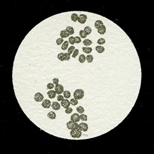
3501897 A colony of Staphylococcus pyogenes after cell division, 1906 (litho) by German School, (20th century); Private Collection; (add.info)

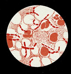
Background imageCellular Collection: Colony of Streptococcus pyogenes, 1906 (litho)

Colony of Streptococcus pyogenes, 1906 (litho)
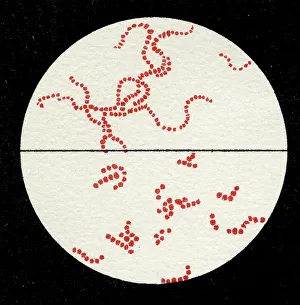
3501894 Colony of Streptococcus pyogenes, 1906 (litho) by German School, (20th century); Private Collection; (add.info.: Bacteria constitute a large domain of prokaryotic microorganisms)

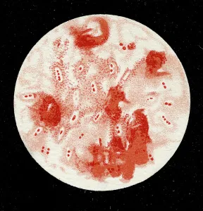
Background imageCellular Collection: Colony of Yersinia pestis, 1906 (litho)

Colony of Yersinia pestis, 1906 (litho)
3501917 Colony of Yersinia pestis, 1906 (litho) by German School, (20th century); Private Collection; (add.info.: Bacteria constitute a large domain of prokaryotic microorganisms)

Background imageCellular Collection: Colony of Salmonella Typhi showing Bacilli with flagella, 1906 (litho)

Colony of Salmonella Typhi showing Bacilli with flagella, 1906 (litho)
3501916 Colony of Salmonella Typhi showing Bacilli with flagella, 1906 (litho) by German School, (20th century); Private Collection; (add.info)

Background imageCellular Collection: Colony of Salmonella typhi, 1906 (litho)

Colony of Salmonella typhi, 1906 (litho)
3501915 Colony of Salmonella typhi, 1906 (litho) by German School, (20th century); Private Collection; (add.info.: Bacteria constitute a large domain of prokaryotic microorganisms)

Background imageCellular Collection: Colony of Bacillus Anthracis in a human spleen, 1906 (litho)

Colony of Bacillus Anthracis in a human spleen, 1906 (litho)
3501905 Colony of Bacillus Anthracis in a human spleen, 1906 (litho) by German School, (20th century); Private Collection; (add.info)


All Professionally Made to Order for Quick Shipping

"Unveiling the Intricacies of Cellular Life: A Journey Through Micrographs" Budding yeast cell: Witness the birth of new life as a budding yeast cell emerges, paving the way for future generations. Nerve and glial cells, light micrograph: Delve into the intricate network of nerve and glial cells that form the foundation of our nervous system. Dicotyledon plant stem, light micrograph: Explore the vibrant world within a dicotyledon plant stem, where cells work tirelessly to support growth and development. Rough endoplasmic reticulum, TEM: Peer into the fascinating realm of rough endoplasmic reticulum through a transmission electron microscope (TEM), revealing its role in protein synthesis. Dendritic cells, artwork: Marvel at an artistic representation showcasing dendritic cells' unique shape and function in immune responses. Cell structure: Uncover the hidden beauty within cellular structures as we unravel their complexity and significance in various biological processes. Mitosis, light micrograph: Observe nature's remarkable spectacle – mitosis – captured under a light microscope; witness how one cell divides into two with precision and grace. Cell division, fluorescent micrograph: Immerse yourself in a mesmerizing display of fluorescent colors capturing another facet of cell division's captivating dance. Dohle bodies in blood cell, micrograph: Discover peculiar "Dohle bodies" within blood cells through high-resolution microscopy—a clue to certain health conditions requiring further investigation. Acute promyelocytic leukemia, micrograph : Gain insight into acute promyelocytic leukemia by examining abnormal cellular characteristics observed under powerful magnification—an invaluable tool for diagnosis and treatment strategies Phagocytosis of fungal spores SEM : Witness nature's defense mechanism unfold as fungal spores are engulfed by cells in a captivating scanning electron microscope (SEM) image.