
Cerebral Collection
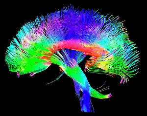
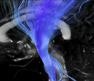
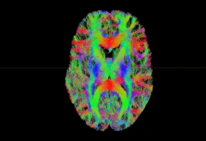
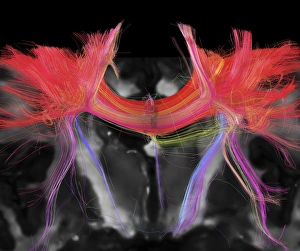
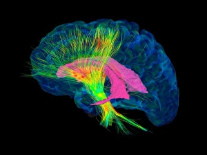
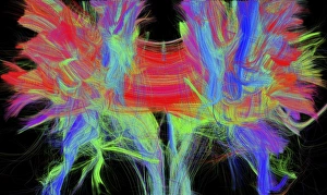
"Cerebral: Unlocking the Mysteries of the Mind" Delve into the intricate web of brain fibres with a DTI MRI scan C017 / 7099


All Professionally Made to Order for Quick Shipping
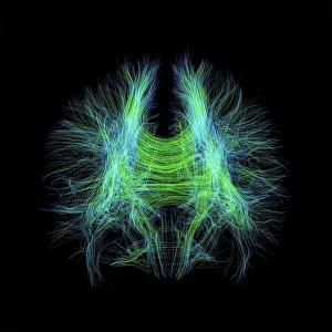
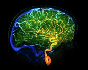
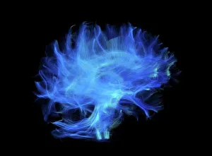
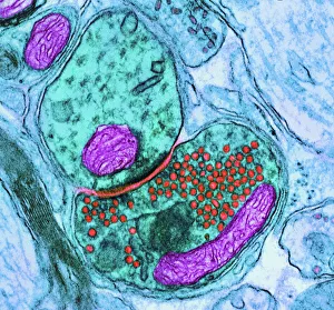
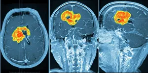
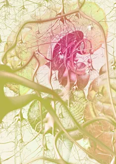
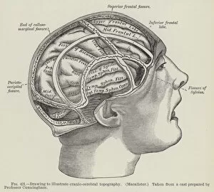
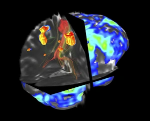
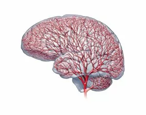
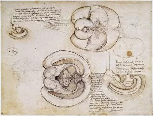
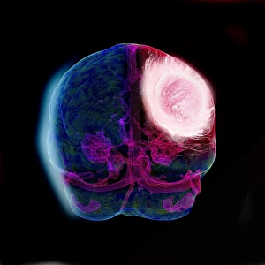
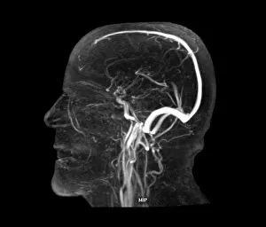
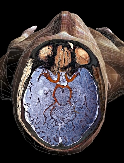
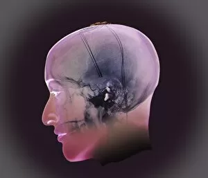
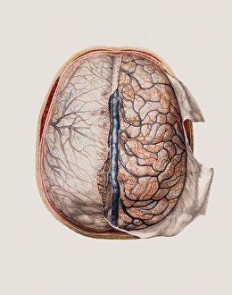
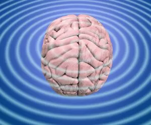
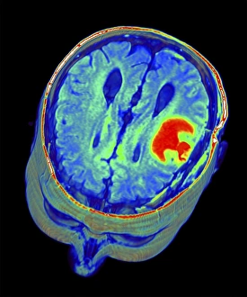
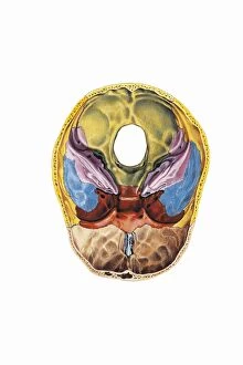
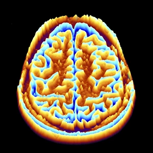
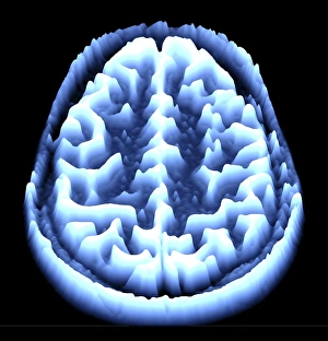
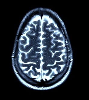
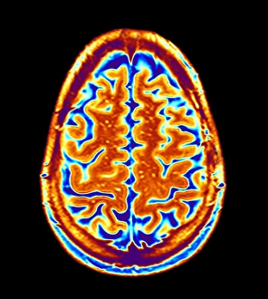
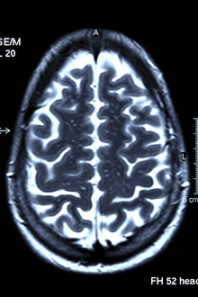
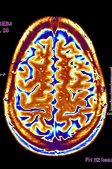
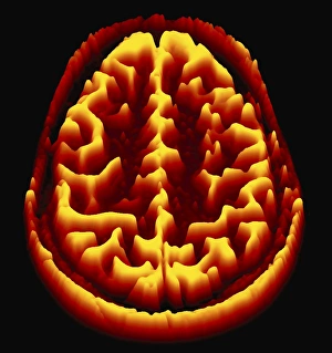
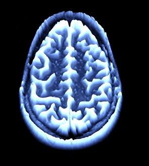
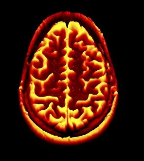
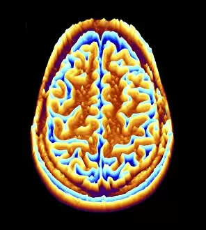
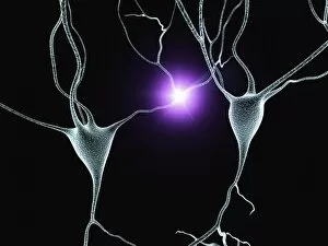
"Cerebral: Unlocking the Mysteries of the Mind" Delve into the intricate web of brain fibres with a DTI MRI scan C017 / 7099, revealing the mesmerizing complexity of our neural pathways. Marvel at the delicate intricacy of brain blood vessels captured in a stunning 3D angiogram C007 / 1981, showcasing their vital role in nourishing our cognitive abilities. Witness synapse nerve junctions through a TEM image, where electrical signals leap across microscopic gaps to facilitate communication within our brains. Explore the challenges posed by a brain tumour as seen in an MRI scan, reminding us of both the resilience and vulnerability inherent in this remarkable organ. Discover how phantom pain after amputation is artistically depicted, shedding light on its enigmatic nature and urging empathy for those who endure it. Uncover the foundations of cognition with insights into skull anatomy and brain structure, providing essential knowledge for understanding our own consciousness. Trace back millions of years to witness primate brain evolution unfold before your eyes - a testament to nature's relentless pursuit of intelligence. Finally, observe cerebral vasculitis through an X-ray image, highlighting potential threats that can disrupt normal blood flow within this crucial system. Intriguing and awe-inspiring, "Cerebral" invites you on an extraordinary journey deep into the recesses of your mind - where wonders await at every turn.