
Choroid Collection
"The Choroid: Unveiling the Artistry of Eye Anatomy" Step into the intricate world of eye anatomy and witness the captivating beauty of the choroid


All Professionally Made to Order for Quick Shipping
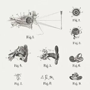
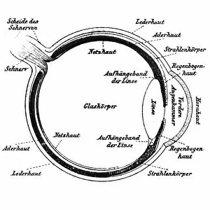
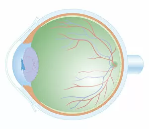
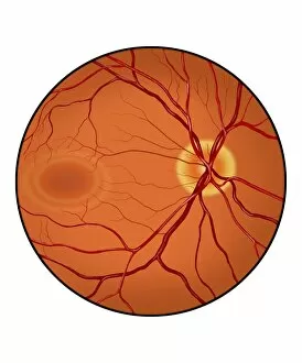
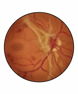
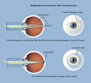
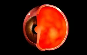
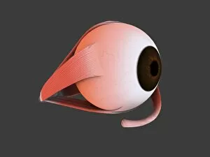
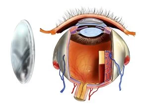
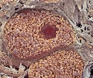
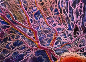
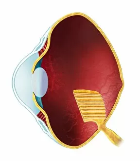
"The Choroid: Unveiling the Artistry of Eye Anatomy" Step into the intricate world of eye anatomy and witness the captivating beauty of the choroid. This vital layer, nestled between the retina and sclera, plays a crucial role in our vision. In one mesmerizing artwork, a scanning electron microscope (SEM) reveals an up-close view of this delicate structure. Its intricate network of blood vessels showcases nature's impeccable design, ensuring nourishment and oxygen supply to the surrounding tissues. Travel back in time as we explore historical illustrations from 1861 and 1898 that depict the anatomy of both human eyes and ears. These masterpieces not only showcase scientific accuracy but also highlight humanity's fascination with unraveling our own complexities. A cross-section biomedical illustration takes us on a journey through every layer within the human eye. The choroid stands out prominently, showcasing its rich pigmentation that absorbs excess light to enhance visual acuity. Conceptual images further deepen our understanding by portraying human eye anatomy alongside skulls or in isolation. These thought-provoking visuals remind us of how intricately connected our vision is to our overall perception and existence. However, amidst this awe-inspiring beauty lies a reminder of vulnerability – macular degeneration affecting the retina. A conceptual image portrays this condition, urging us to appreciate each moment when our sight remains unimpaired. Lastly, an orbital cut unravels more secrets as it exposes nerves like abducent nerve with ciliary ganglion and oculomotor nerve. Such imagery reminds us that behind every blink or gaze lies an orchestra conducted by these microscopic marvels. The choroid invites us into its realm where science meets artistry.