
Ciliates Collection
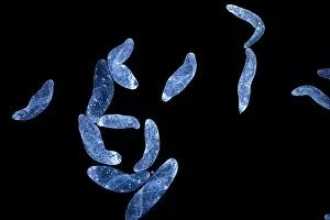
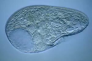
"Ciliates: Fascinating Microscopic Creatures Revealed in Stunning Detail" Step into the mesmerizing world of ciliates, as captured by powerful light micrographs


All Professionally Made to Order for Quick Shipping
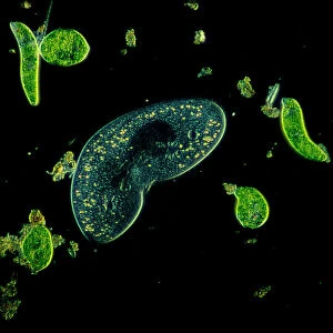
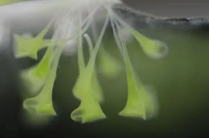
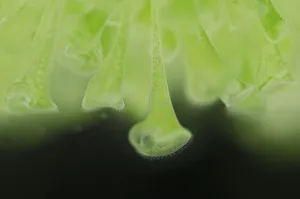
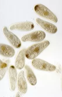
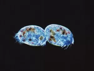
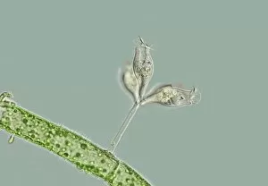
"Ciliates: Fascinating Microscopic Creatures Revealed in Stunning Detail" Step into the mesmerizing world of ciliates, as captured by powerful light micrographs. These protozoans, with their distinct kidney-shaped bodies, are surrounded by a vibrant array of Euglena sp. , creating a breathtaking sight under 900x magnification. The intricate details and vivid colors showcased in this Ciliata lithograph from 1899-1904 transport us to an era when scientific exploration was at its peak. In another captivating image, we witness the symbiotic relationship between trumpet animalcules (Stentor polymorphus) and algal symbionts (Chlorella). Found across Europe, these tiny organisms form a harmonious partnership where Stentor provides shelter while Chlorella contributes essential nutrients. Picture No. 11014599 beautifully captures this delicate alliance that thrives amidst nature's wonders. As our journey continues through the microscopic realm, we encounter more enchanting scenes like Picture No. 11675491, Picture No. 11675490, Picture No. 11675489, and Picture No. 11675488 – each unveiling unique aspects of ciliate life that leave us awe-inspired. Amongst them is Chilodonella ciliate protozoa illuminated by LM; its graceful presence showcases the elegance hidden within even the tiniest creatures on Earth. Spirostomum protozoa C017/8347 captivates with its elongated body and undulating movements—a testament to nature's boundless creativity. Finally, Blepharisma protozoan graces us with its presence in a stunning light micrograph—an organism so intricately designed that it seems almost otherworldly. These glimpses into the world remind us of the vast diversity found within our own planet—often unseen but ever-present beneath our feet or floating in nearby waters.