
Contrast Medium Collection
"Exploring the Intricacies of the Human Body: The Power of Contrast Medium" In the realm of medical imaging


All Professionally Made to Order for Quick Shipping
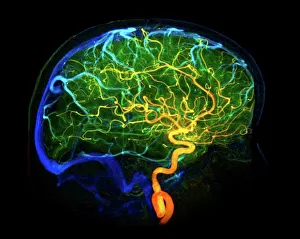
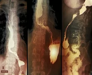
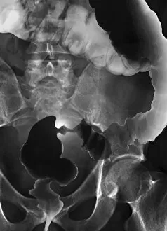
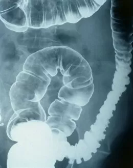
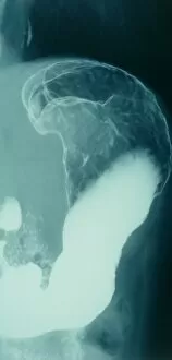
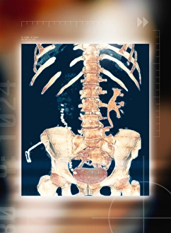
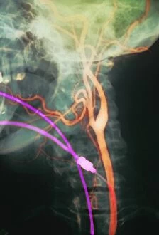
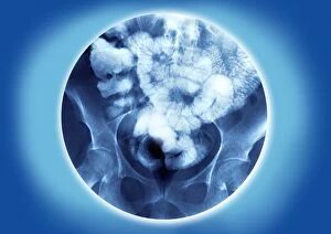
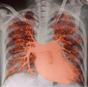
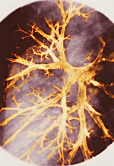
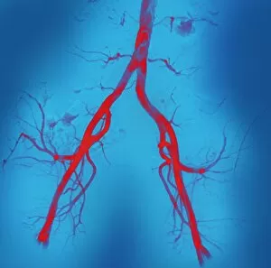
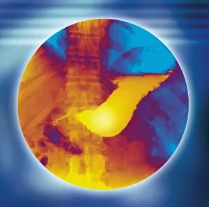
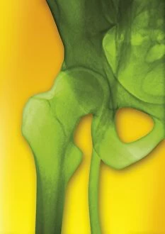
"Exploring the Intricacies of the Human Body: The Power of Contrast Medium" In the realm of medical imaging, contrast medium serves as a remarkable tool to shed light on various conditions and anomalies within our bodies. From brain blood vessels captured in a mesmerizing 3D angiogram C007/1981 to neck and shoulder arteries revealed through X-ray technology, these contrasting agents allow us to delve deeper into the intricate network that sustains our lives. Venturing beyond conventional examinations, contrast medium unravels mysteries such as elephantiasis, exposing its severity through an enlightening X-ray image. Similarly, throat cancer comes under scrutiny with detailed X-rays that aid in diagnosis and treatment decisions. But it doesn't stop there; contrast medium unveils more secrets hidden beneath the surface. A full bladder is brought into focus through X-ray cystography, providing valuable insights for urological assessments. Colon cancer finds itself exposed by barium-enhanced X-rays (C018/0574), while colonic spasms are captured in their dynamic essence using this same technique. The journey continues with stomach cancer being unveiled via barium-infused X-rays – a powerful diagnostic tool enabling early detection and intervention. Meanwhile, female reproductive organs come alive under the gaze of radiographic images (C015/6051), offering invaluable information for gynecological evaluations. Lastly, we explore the small intestine's complex terrain using barium-enhanced X-rays – an illuminating experience that aids in diagnosing gastrointestinal disorders. Contrast medium acts as a guiding light for medical professionals worldwide - revealing what was once invisible or elusive within our bodies. Its ability to enhance visibility empowers healthcare providers to make informed decisions and provide effective treatments tailored specifically to each patient's needs. As technology advances further, so too does our understanding of human anatomy - all thanks to this extraordinary substance known as contrast medium.