Fraud Blocker Skip to main content

Cranium Collection

"Cranium: Unveiling the Mysteries of the Human Skull and Beyond" Delving into the depths of human anatomy

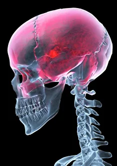
Background imageCranium Collection: Headache, X-ray artwork

Headache, X-ray artwork
Headache. Computer X-ray artwork of a lateral view of a human skull. The red area represents pain caused by a headache (cephalalgia)
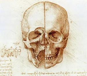
Background imageCranium Collection: Skull anatomy by Leonardo da Vinci

Skull anatomy by Leonardo da Vinci. Historical artwork and notes on the anatomy of the human skull and teeth, by the Italian artist and scientist Leonardo da Vinci (1452-1519)

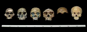
Background imageCranium Collection: Hominid crania

Hominid crania
L to R: Australopithecus africanus; Homo rudolfensis; H.erectus; H. heildebergensis; H. neanderthalensis and H. sapiens. Arranged in chronological order these specimens (casts)

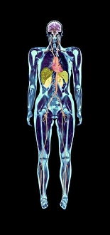
Background imageCranium Collection: Full body scan, MRI scan

Full body scan, MRI scan
Full body scan of a female swimmer. Her brain is purple (top), her heart and arteries are red/orange (upper centre), her kidneys are pale green (centre)

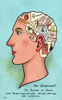
Background imageCranium Collection: Satire on Phrenology

Satire on Phrenology
Satire on the study of Phrenology - a Free Knowledgey - illustrating the worst elements of human behavious illustrated in different zones of the head (including Obscene Language, criminal activity)

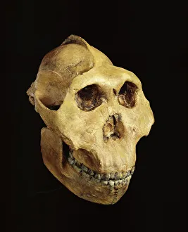
Background imageCranium Collection: Paranthropus boisei (Zinjanthropus) cranium (OH5)

Paranthropus boisei (Zinjanthropus) cranium (OH5)
Cast of the cranium of a young male of Paranthropus boisei discovered by Mary Leakey in 1959 at Olduvai Gorge. The specimen which is the Holotype of Zinjanthropus boisei (Leakey 1959)

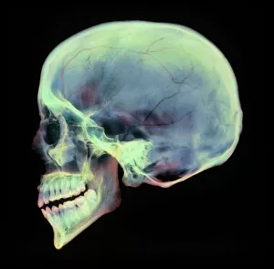
Background imageCranium Collection: Human skull, X-ray

Human skull, X-ray
Human skull. Coloured X-ray of a sagittal section through a human skull. The skull has been sliced in half down the centre, revealing details of the internal structures

Background imageCranium Collection: Horse skull

Horse skull (Equus caballus), side view X-ray

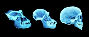
Background imageCranium Collection: Primate skulls

Primate skulls. Coloured x-rays of the skulls of a gorilla (Gorilla gorilla, left), chimpanzee (Pan trogoldytes, centre) and human (Homo sapien, left)

Background imageCranium Collection: Sahelanthropus tchadensis skull

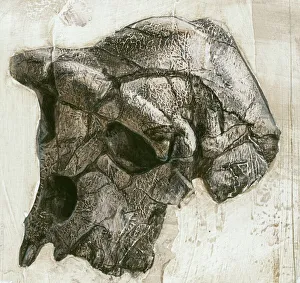
Sahelanthropus tchadensis skull. Artwork of a reconstruction of the Toumai skull, one of only a small number of fossils of the hominin Sahelanthropus tchadensis

Background imageCranium Collection: Human skull

Human skull
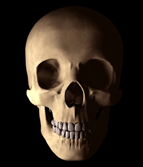
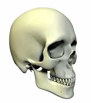
Skull. Computer artwork of a healthy human skull seen from the front. The bones of the cranium (upper centre) are fused together to form a solid structure that encloses and protects the brain

Background imageCranium Collection: Homo erectus, Java Man cranium (Sangiran 17) cast

Homo erectus, Java Man cranium (Sangiran 17) cast
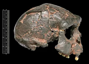
Lateral view of partially reconstructed cranium of Homo erectus Java Man about 700, 000 years old known as Sangiran 17. Discovered by Towikromo in 1969

Background imageCranium Collection: Skull anatomy

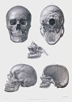
Skull anatomy. Historical anatomical artwork of various views of the human skull. The frontal view (upper left) shows the teeth, nose and eye spaces

Background imageCranium Collection: Skull and Crossbones - Inverted

Skull and Crossbones - Inverted
The Jolly Roger is the name given to any of various flags flown to identify a ships crew as pirates. The flag most usually identified as the Jolly Roger today is the skull and crossbones

Background imageCranium Collection: Skull and Crossbones

Skull and Crossbones
The Jolly Roger is the name given to any of various flags flown to identify a ships crew as pirates. The flag most usually identified as the Jolly Roger today is the skull and crossbones

Background imageCranium Collection: Homo sp. skulls C016 / 5933

Homo sp. skulls C016 / 5933
Side views of Homo erectus (Sangiran), H. heidelbergensis (Broken Hill), H. neanderthalensis, (La Ferrassie) and H. sapiens (Polynesia) skulls. Natural History Museum, London, UK

Background imageCranium Collection: Skull of Neanderthal (Homo neanderthalensis)

Skull of Neanderthal (Homo neanderthalensis). Krapina. Croatia. Natural History Museum. London. United Kingdom

Background imageCranium Collection: Allosaurus cranium

Allosaurus cranium
A detail of the skull of Allosaurus, the Upper Jurassic carnivorous dinosaur that lived 153 to 135 million years ago. On display at the Natural History Museum, London

Background imageCranium Collection: Skull

Skull. Computer artwork of an oblique view of a human skull. The bones of the cranium are fused together to form a solid structure that encloses and protects the brain

Background imageCranium Collection: Phrenology bust

Phrenology bust. Phrenology was a popular pseudoscience of the nineteenth century. The basis for the theory lay in a misplaced belief that a persons character

Background imageCranium Collection: Homo erectus skull

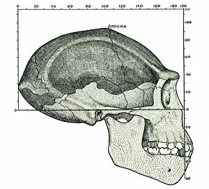
Homo erectus skull. Artwork, from 1931, showing a reconstruction of the skull of Java Man, based on the fossil skull fragments (dark grey) discovered by Dubois in Java in 1891

Background imageCranium Collection: Homo erectus cranium C013 / 6552

Homo erectus cranium C013 / 6552
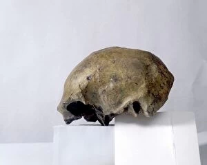
Cast of a Homo erectus cranium. The fossil this cast was taken from is known as Solo Man (Ngandong 1), and was found at Ngandong, close to the Solo River on the island of Java, Indonesia

Background imageCranium Collection: Cranium 1-4 and part of sternum 5,6 of the extinct Rodrigues solitaire

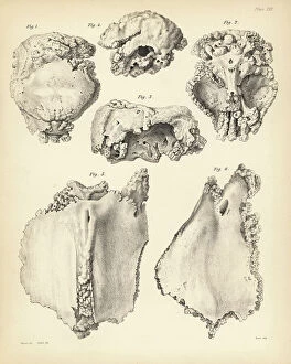
Cranium 1-4 and part of sternum 5,6 of the extinct Rodrigues solitaire. Lithograph by Joseph Dinkel after Werner
FLO4612182 Cranium 1-4 and part of sternum 5,6 of the extinct Rodrigues solitaire. Lithograph by Joseph Dinkel after Werner from Hugh Edwin Strickland and Alexander Gordon Melville's The Dodo

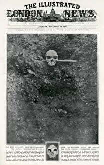
Background imageCranium Collection: Diagram of the lead and zinc mine in Broken Hill, Northern Rhodesia (now Kabwe, Zambia)

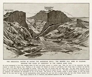
Diagram of the lead and zinc mine in Broken Hill, Northern Rhodesia (now Kabwe, Zambia) - the site of the discovery of a Cranium of Homo rhodesiensis, in Mutwe Wa Nsofu Area on June 17

Background imageCranium Collection: Photograph of the lead and zinc mine in Broken Hill, Northern Rhodesia (now Kabwe

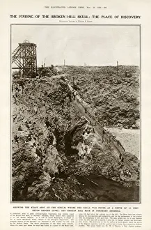
Photograph of the lead and zinc mine in Broken Hill, Northern Rhodesia (now Kabwe, Zambia) - the site of the discovery of a Cranium of Homo rhodesiensis, in Mutwe Wa Nsofu Area on June 17

Background imageCranium Collection: Anatomy of human skull from different angles

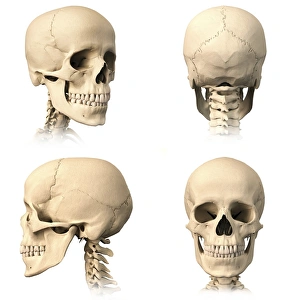
Anatomy of human skull from different angles

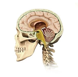
Background imageCranium Collection: Human head anatomy, artwork

Human head anatomy, artwork
Human head anatomy, computer artwork

Background imageCranium Collection: Cranium and sternum of the extinct Rodrigues

Cranium and sternum of the extinct Rodrigues
Cranium 1-4 and part of sternum 5, 6 of the extinct Rodrigues solitaire, Pezophaps solitaria, in the Parisian Collection. Lithograph by Joseph Dinkel after Werner from Hugh Edwin Strickland

Background imageCranium Collection: Cranium of Homo rhodesiensis, discovered in Mutwe Wa Nsofu Area in a lead and zinc

Cranium of Homo rhodesiensis, discovered in Mutwe Wa Nsofu Area in a lead and zinc mine in Broken Hill, Northern Rhodesia (now Kabwe, Zambia) on June 17, 1921 by Tom Zwiglaar, a Swiss miner. H

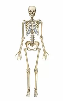
Background imageCranium Collection: Human skeletal system, front view

Human skeletal system, front view

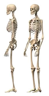
Background imageCranium Collection: Anatomy of male human skeleton, side view and perspective view

Anatomy of male human skeleton, side view and perspective view

Background imageCranium Collection: Egyptian skulls

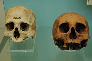
Egyptian skulls
Egypt. Skulls. Initial Late Period. One of them is dated after 664 BC. 26th dynasty or Saite Period (672-525 BC). From cemetery near Tura. The other has uncertain date and unknown origin

Background imageCranium Collection: Egyptian skull

Egyptian skull. British Museum. London. United Kingdom

Background imageCranium Collection: St. Jerome. First half of the 18th century. Byzantine Museum

St. Jerome. First half of the 18th century. Byzantine Museum. Zante. Ionian Islands. Greece

Background imageCranium Collection: Concepts: reincarnation, an allegory

Concepts: reincarnation, an allegory
An allegory of reincarnation-the infant is reborn from the dead self. Date: 1907

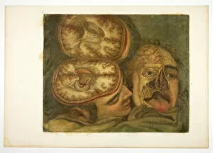
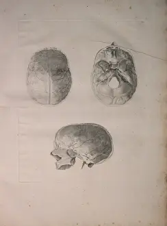
Background imageCranium Collection: Cranial Dissection, plate five from Anatomy of the Head, in Printed Paintings, 1748

Cranial Dissection, plate five from Anatomy of the Head, in Printed Paintings, 1748

Background imageCranium Collection: Henri de Mondeville teaching anatomy, University of Montepellier, 1312

Henri de Mondeville teaching anatomy, University of Montepellier, 1312, illustration from Histoire de la Nation
STC397223 Henri de Mondeville teaching anatomy, University of Montepellier, 1312, illustration from Histoire de la Nation Francaise, Sciences, Volumes I & II, by Gabriel Hanutaux (1853-1944)

Background imageCranium Collection: Albinus II, Pl. III, Cranial bone, illustration from Tabulae ossium humanorum

Albinus II, Pl. III, Cranial bone, illustration from Tabulae ossium humanorum
HMB306101 Albinus II, Pl. III, Cranial bone, illustration from Tabulae ossium humanorum, by Bernhard Siegfried Albinus (1697-1770), published by J.&H. Verbeek, bibliop

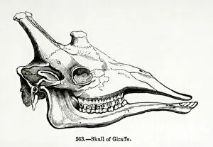
Background imageCranium Collection: Skull of a Giraffe (litho) (b / w photo)

Skull of a Giraffe (litho) (b / w photo)
XJF275536 Skull of a Giraffe (litho) (b/w photo) by English School; Private Collection; English, out of copyright

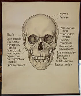
Background imageCranium Collection: Poster of a human skull (litho)

Poster of a human skull (litho)
HMB306165 Poster of a human skull (litho) by German School; 126x106 cm; Humboldt-Universitaet, Berlin, Germany; (add.info.: Wandtafel, Mensch, Schaedel von vorn; ); eHumboldt Innovation

Background imageCranium Collection: Craniological map of the head and the compression cap, caricature of phrenology

Craniological map of the head and the compression cap, caricature of phrenology
CHT164121 Craniological map of the head and the compression cap, caricature of phrenology (engraving) (b/w photo) by French School, (19th century); Musee d Histoire de la Medecine, Paris

Background imageCranium Collection: Albinus II, Pl. I, illustration from Tabulae ossium humanorum

Albinus II, Pl. I, illustration from Tabulae ossium humanorum
HMB306100 Albinus II, Pl. I, illustration from Tabulae ossium humanorum, by Bernhard Siegfried Albinus (1697-1770), 1729, published by J.&H. Verbeek, bibliop

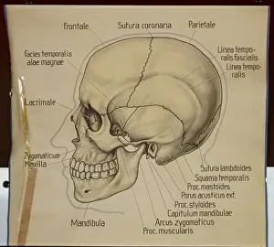
Background imageCranium Collection: Poster of the side view of a human skull (litho)

Poster of the side view of a human skull (litho)
HMB306164 Poster of the side view of a human skull (litho) by German School; 130x125 cm; Humboldt-Universitaet, Berlin, Germany; (add.info.: Wandtafel, Mensch)

Background imageCranium Collection: Ms Lat. 7134 fol. 1 Trepanation, from Chirugie de Maitre Rolandi (vellum)

Ms Lat. 7134 fol. 1 Trepanation, from Chirugie de Maitre Rolandi (vellum)
CHT179389 Ms Lat. 7134 fol.1 Trepanation, from Chirugie de Maitre Rolandi (vellum) by French School, (14th century); Bibliotheque Nationale, Paris, France; eArchives Charmet; French, out of copyright

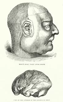
Background imageCranium Collection: Swifts Head, taken after death, and cast of the interior of Swifts cranium

Swifts Head, taken after death, and cast of the interior of Swifts cranium
1772349 Swifts Head, taken after death, and cast of the interior of Swifts cranium, from Gullivers Travels, published c.1875 (litho) by English School

Background imageCranium Collection: Occipital bone, outer surface (engraving)

Occipital bone, outer surface (engraving)
3634213 Occipital bone, outer surface (engraving) by English School, (19th century); Private Collection; (add.info.: Occipital bone, outer surface)

Background imageCranium Collection: Illustration for life of Jonathan Swift (engraving)

Illustration for life of Jonathan Swift (engraving)
5989453 Illustration for life of Jonathan Swift (engraving) by Morten, Thomas (1836-66); Private Collection; (add.info.: Swifts head, taken after death; cast of the interior of the cranium of Swift)

Background imageCranium Collection: Albinus II, Pl. II, Skull, illustration from Tabulae ossium humanorum

Albinus II, Pl. II, Skull, illustration from Tabulae ossium humanorum
HMB306102 Albinus II, Pl. II, Skull, illustration from Tabulae ossium humanorum, by Bernhard Siegfried Albinus (1697-1770), published by J.&H. Verbeek, bibliop


All Professionally Made to Order for Quick Shipping

"Cranium: Unveiling the Mysteries of the Human Skull and Beyond" Delving into the depths of human anatomy, Leonardo da Vinci's intricate sketches of skull anatomy offer a glimpse into his fascination with unraveling the secrets held within our craniums. From his detailed drawings to modern-day advancements, we continue to explore this enigmatic structure. Hominid crania provide us with a window into our evolutionary past, showcasing how our ancestors' skulls have evolved over time. With full-body scans and MRI technology, scientists can now peer inside these ancient relics, uncovering clues about their lifestyles and adaptations. In a satirical twist on phrenology - an outdated pseudoscience that claimed personality traits could be determined by skull shape - we find humor in our quest for understanding. Yet amidst the satire lies an appreciation for how far we've come in deciphering the complexities of cranial morphology. The Paranthropus boisei (Zinjanthropus) cranium (OH5) takes us back millions of years, offering insights into early hominids' robust features. Meanwhile, X-ray images reveal intricate details of both human and animal skulls – from horses to primates – highlighting similarities and differences across species. One cannot ignore Sahelanthropus tchadensis; its fossilized skull provides a crucial link between apes and humans. This remarkable discovery challenges previous notions about our origins while fueling further exploration into humanity's beginnings. But it is not just scientific curiosity that draws attention to the cranium; it also holds significance in everyday life. Headaches become tangible through X-ray artwork, capturing both pain and beauty simultaneously. The human skull itself serves as a reminder of mortality but also showcases resilience throughout history. Finally, Homo erectus' Java Man cranium (Sangiran 17) cast allows us to step back in time once more – witnessing glimpses of ancient lives and pondering the mysteries that still surround our own existence.