
Cytology Collection
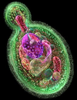
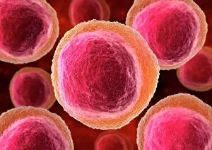
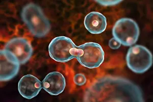
Cytology: Exploring the Intricate World of Cells Budding yeast cell: Witness the remarkable process of cellular reproduction as a budding yeast cell emerges


All Professionally Made to Order for Quick Shipping
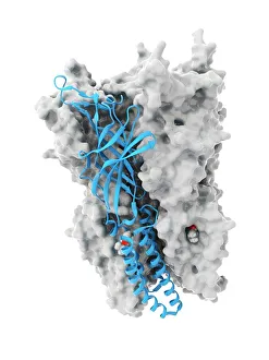
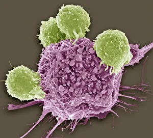
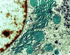
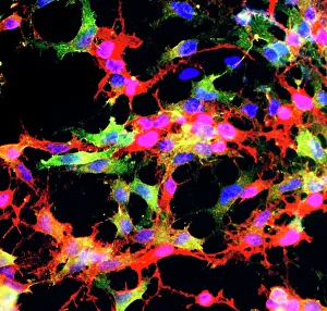
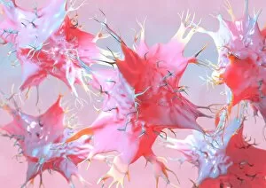
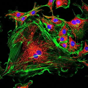
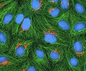
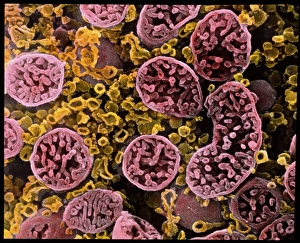
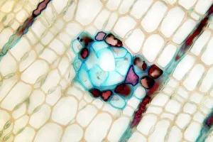
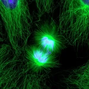
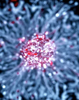
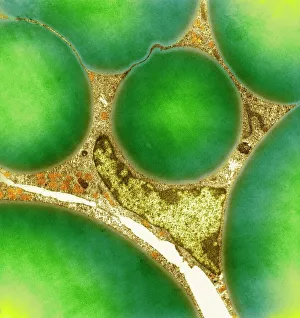
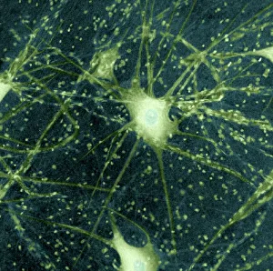
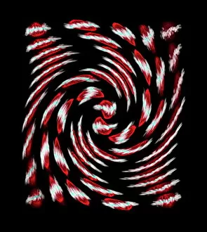
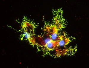
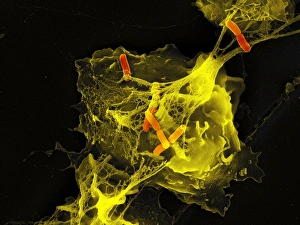
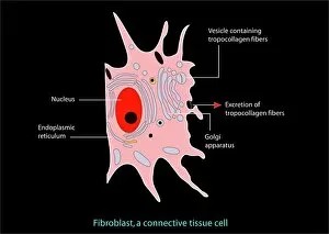
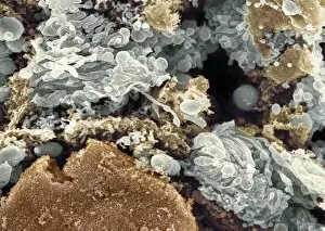
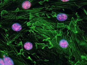
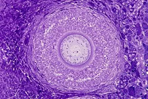
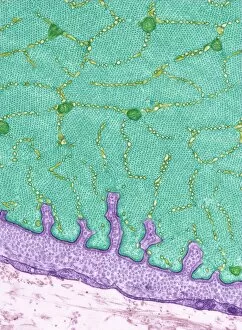
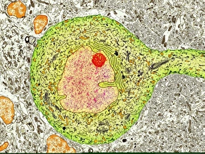
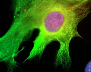
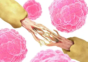
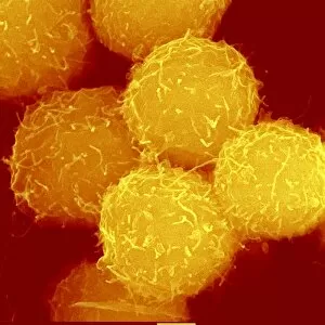
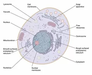
Cytology: Exploring the Intricate World of Cells Budding yeast cell: Witness the remarkable process of cellular reproduction as a budding yeast cell emerges, paving the way for new life. T lymphocytes and cancer cell, SEM C001 / 1679: Delve into the battle between our immune system's T lymphocytes and cancer cells, captured in stunning detail through scanning electron microscopy. Anaesthetic inhibiting an ion channel C015 / 6718: Uncover the secrets behind anesthesia as we observe its impact on ion channels within cells, revealing how it alters our perception of pain. Glial cells, confocal light micrograph: Immerse yourself in the intricate network of glial cells that support and protect neurons in our nervous system, visualized using confocal light microscopy. HeLa cells, light micrograph C017 / 8299: Explore one of the most famous cell lines - HeLa cells - under a microscope to gain insights into their structure and function within biomedical research. Rough endoplasmic reticulum, TEM: Journey deep inside a cell to witness the rough endoplasmic reticulum at work – a vital organelle involved in protein synthesis – revealed through transmission electron microscopy. Glial stem cell culture, light micrograph: Step into the realm of regenerative medicine as we observe glial stem cells cultured in vitro with hopes of unlocking their potential for repairing damaged neural tissue. Dendritic cells, artwork: Marvel at an artistic representation showcasing dendritic cells' crucial role as sentinels of our immune system – capturing antigens and presenting them to other immune cells for recognition and response. Cell structure: Embark on a visual journey exploring various cellular structures that form the building blocks of life itself – from membranes to cytoplasm to nuclei – highlighting their diverse functions.