
Diatom Collection
Diatoms, the mesmerizing art of nature's microscopic algae, captivate our imagination with their intricate beauty


All Professionally Made to Order for Quick Shipping
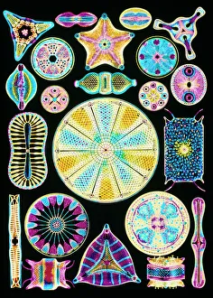
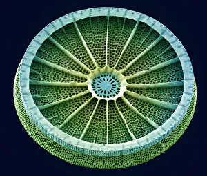
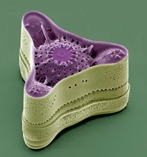
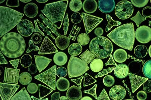
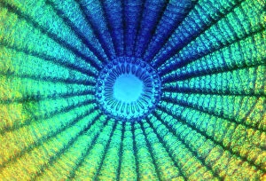
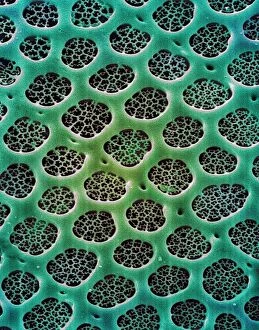
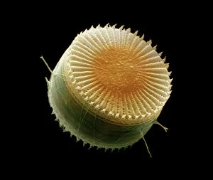
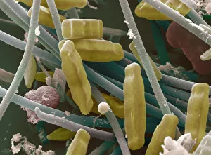
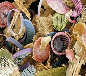
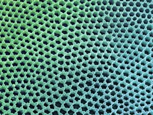
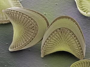
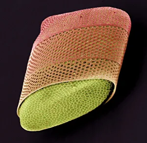
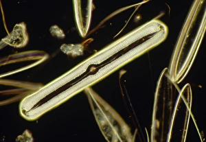
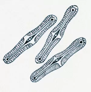
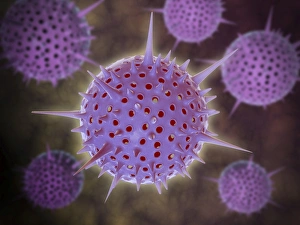
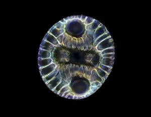
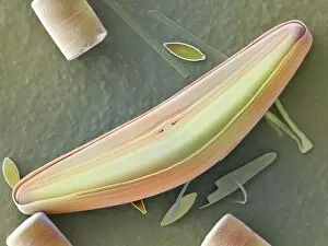
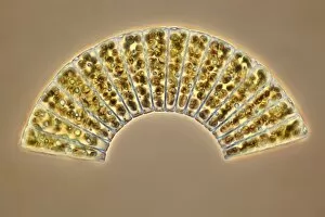
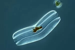
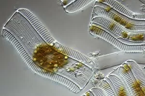
Diatoms, the mesmerizing art of nature's microscopic algae, captivate our imagination with their intricate beauty. Ernst Haeckel's stunning illustrations bring these diatom masterpieces to life, showcasing their delicate and symmetrical forms. Through scanning electron microscopy (SEM), we delve into a world unseen by the naked eye, where diatoms reveal their astonishing complexity. From marine plankton samples, diatoms emerge as tiny wonders of nature. SEM allows us to explore their structures up close and personal – each image revealing a new dimension of their elegance. The sheer diversity within this single group is astounding; every species possesses its own unique pattern and design. As we peer through the lens of SEM, we witness diatoms in all their glory. Their exquisitely detailed cell walls become apparent, displaying an array of shapes and textures that resemble miniature works of art. These microorganisms have mastered the artistry of survival in aquatic environments worldwide. Even in fossilized form, diatoms continue to amaze us with their preserved beauty under SEM examination. These ancient remnants offer glimpses into past ecosystems while reminding us of the enduring legacy left by these remarkable organisms. In this captivating collection of images captured through SEM technology, we are reminded that even the tiniest inhabitants on Earth possess extraordinary intricacy worth exploring. Diatoms stand as a testament to both the artistic genius found within nature and its ability to create breathtaking marvels at any scale imaginable.