
Dilated Collection
"Dilated: Exploring the Intricacies of Varicose Veins and Beyond" Varicose veins, often referred to as dilated blood vessels


All Professionally Made to Order for Quick Shipping
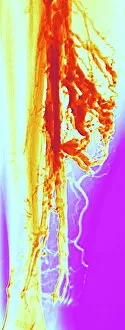
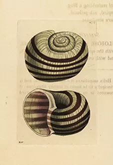
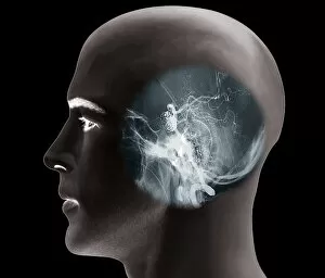
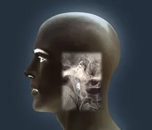
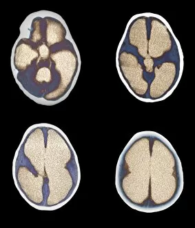
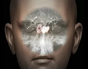
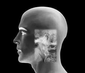
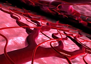
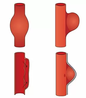
"Dilated: Exploring the Intricacies of Varicose Veins and Beyond" Varicose veins, often referred to as dilated blood vessels, can cause discomfort and aesthetic concerns for many individuals. X-ray imaging allows medical professionals to visualize the extent of varicose veins and develop appropriate treatment plans. The Pila ampullacea, a species of freshwater snail with a distinctive dilated shell, showcases nature's unique adaptations. Kennedya dilatata, commonly known as Dilated Kennedya, is a vibrant flowering plant native to Australia that adds beauty to any garden. Anthony Trollope's illustration for Barchester Towers depicts characters in an emotionally diluted state, highlighting their complex relationships. Lung disease can be detected through X-ray scans like C017 / 7568, enabling early diagnosis and intervention for better patient outcomes. Abdominal aortic aneurysm is characterized by the dilation of the main blood vessel supplying oxygen-rich blood to the abdomen; ultrasound scans like C017 / 7649 aid in its detection. Aneurysm treatment often involves angiograms (such as C018 / 0537 and C018 / 0536), which use contrast dye injected into blood vessels to identify areas of dilation requiring intervention. Hydrocephalus is a condition where excess cerebrospinal fluid accumulates within the brain's ventricles; CT scans (C018 / 0568) help diagnose this condition accurately. Aneurysm treatment continues to evolve with advancements in medical technology; angiograms (like C018 / 0525) play a crucial role in guiding interventions for patients' well-being. Hydrocephalus affects individuals differently; CT scans (C018 / 0567) provide valuable insights into this condition's progression and inform personalized treatment plans.