Fraud Blocker Skip to main content

Dissected Collection

"Unveiling the Intricacies of Life: A Journey through Dissected Hints" Step into the world of Leonardo da Vinci's skull anatomy

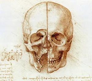
Background imageDissected Collection: Skull anatomy by Leonardo da Vinci

Skull anatomy by Leonardo da Vinci. Historical artwork and notes on the anatomy of the human skull and teeth, by the Italian artist and scientist Leonardo da Vinci (1452-1519)

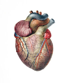
Background imageDissected Collection: Heart

Heart. Historical anatomical artwork of the human heart, seen from the front. Coronary blood vessels are seen on the surface of the heart

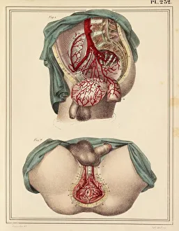
Background imageDissected Collection: Male groin arteries, 1825 artwork

Male groin arteries, 1825 artwork
Male groin arteries. Dissections of a male groin to show areas supplied by the internal iliac artery (red) and its branches
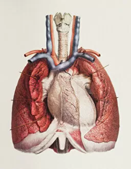
Background imageDissected Collection: Heart and lungs

Heart and lungs. Historical anatomical artwork of the human heart and lungs, seen from the front. Dissection hooks have been used to draw back the lungs (red)
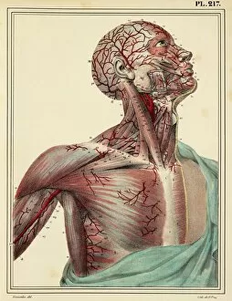
Background imageDissected Collection: Head and chest arteries, 1825 artwork

Head and chest arteries, 1825 artwork
Head and chest arteries. Superficial arteries (red) of the chest, neck and head, which are visible when the skin and muscle fascia are removed

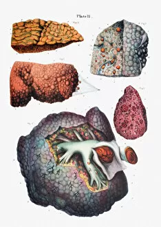
Background imageDissected Collection: Liver cirrhosis

Liver cirrhosis. Artwork taken from Sir Robert Carswells textbook Pathological Anatomy, published 1838. Cirrhosis is a disease in which the liver tissue is replaced with scar tissue

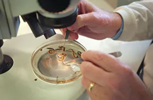
Background imageDissected Collection: Scientist working with a ragworm specimen

Scientist working with a ragworm specimen
Scientist dissecting a ragworm specimen, held at the Natural History Museum, London

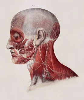
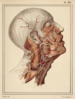
Background imageDissected Collection: Facial nerves

Facial nerves. Historical anatomical artwork of a side view of a dissected human head showing the nerves (white) and muscles (red) of the face and neck

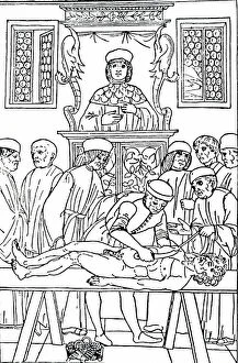
Background imageDissected Collection: An anatomy lecture at Padua

An anatomy lecture at Padua
5311884 An anatomy lecture at Padua; (add.info.: Woodcut engraving of an anatomy lecture at Padua. The professor remains in his chair reading from a book)

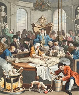
Background imageDissected Collection: The Reward of Cruelty, from The Four Stages of Cruelty, illustration from Hogarth Restored

The Reward of Cruelty, from The Four Stages of Cruelty, illustration from Hogarth Restored
STC451184 The Reward of Cruelty, from The Four Stages of Cruelty, illustration from Hogarth Restored: The Whole Works of the celebrated William Hogarth, re-engraved by Thomas Cook, pub

Background imageDissected Collection: Ms Hunter 364 Table I Dissection instruments, from Anatomical Tables

Ms Hunter 364 Table I Dissection instruments, from Anatomical Tables, by John Banister (1540-1610) (oil on brown paper)
GUL174528 Ms Hunter 364 Table I Dissection instruments, from Anatomical Tables, by John Banister (1540-1610) (oil on brown paper) by English School, (16th century); Glasgow University Library

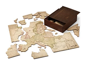
Background imageDissected Collection: Europe divided into its Kingdoms, c. 1766 (hand-coloured engraved sheet mounted on wood)

Europe divided into its Kingdoms, c. 1766 (hand-coloured engraved sheet mounted on wood)
6244298 Europe divided into its Kingdoms, c.1766 (hand-coloured engraved sheet mounted on wood) by Spilsbury, John (1739-69); 453x480 cm; Private Collection; (add.info)

Background imageDissected Collection: Page from The Pictorial Museum of Animated Nature (engraving)

Page from The Pictorial Museum of Animated Nature (engraving)
979869 Page from The Pictorial Museum of Animated Nature (engraving) by English School, (19th century); Private Collection; (add.info)

Background imageDissected Collection: Botanical Dissection: Sour Cherry (Prunus cerasus), c. 1778-90 (w / c & graphite on paper)

Botanical Dissection: Sour Cherry (Prunus cerasus), c. 1778-90 (w / c & graphite on paper)
880315 Botanical Dissection: Sour Cherry (Prunus cerasus), c. 1778-90 (w/c & graphite on paper) by RedoutA©, Pierre-Joseph (1759-1840); Minneapolis Institute of Arts, MN

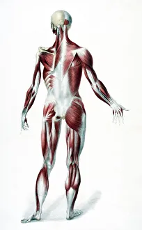
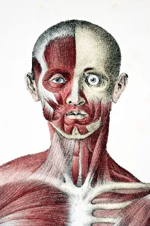
Background imageDissected Collection: Dissected plate of the human body (colour litho)

Dissected plate of the human body (colour litho)
2799323 Dissected plate of the human body (colour litho) by English School, (19th century); Private Collection; (add.info.: Dissected plate of the human body)

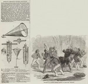
Background imageDissected Collection: Russian Submarine Infernal Machines (engraving)

Russian Submarine Infernal Machines (engraving)
1608054 Russian Submarine Infernal Machines (engraving) by English School, (19th century); Private Collection; (add.info.: Russian Submarine Infernal Machines)

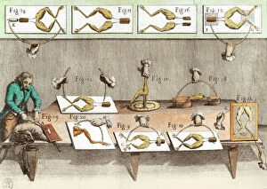
Background imageDissected Collection: Experiment on frogs by Louis Galvani (1737-98) late 18th century (engraving) (b / w photo)

Experiment on frogs by Louis Galvani (1737-98) late 18th century (engraving) (b / w photo)
XIR217898 Experiment on frogs by Louis Galvani (1737-98) late 18th century (engraving) (b/w photo) by French School, (18th century); Private Collection; (add.info.: experiment on frogs; ); French

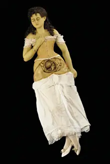
Background imageDissected Collection: Anatomical model of a pregnant woman (wax & textiles)

Anatomical model of a pregnant woman (wax & textiles)
1123883 Anatomical model of a pregnant woman (wax & textiles) by English School, (19th century); Private Collection; (add.info)

Background imageDissected Collection: Experiment on frogs, illustration from De viribus electricitatis in motu

Experiment on frogs, illustration from De viribus electricitatis in motu
XIR217897 Experiment on frogs, illustration from De viribus electricitatis in motu muscumari (engraving) (b/w photo) by French School, (18th century); Bibliotheque Nationale, Paris

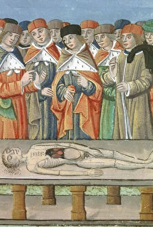
Background imageDissected Collection: Ms Hunter 9 f. 36r Dissection, from De Medina Fen I

Ms Hunter 9 f. 36r Dissection, from De Medina Fen I
GUL174578 Ms Hunter 9 f.36r Dissection, from De Medina Fen I. II, by Avicenna (980-1037) (vellum) by Flemish School, (15th century); 41.3x29.8 cm; Glasgow University Library

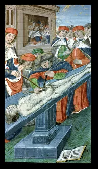
Background imageDissected Collection: Ms Hunter 9 30v Dissection, from De Medina Fen I. II

Ms Hunter 9 30v Dissection, from De Medina Fen I. II
GUL174576 Ms Hunter 9 30v Dissection, from De Medina Fen I. II, by Avicenna (980-1037) (vellum) by Flemish School, (15th century); 41.3x29.8 cm; Glasgow University Library

Background imageDissected Collection: Ms Hunter 9 10r Dissection, from De Medina Fen I. II

Ms Hunter 9 10r Dissection, from De Medina Fen I. II
GUL174573 Ms Hunter 9 10r Dissection, from De Medina Fen I. II, by Avicenna (980-1037) (vellum) by Flemish School, (15th century); 41.3x29.8 cm; Glasgow University Library

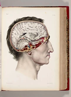
Background imageDissected Collection: A hand coloured lithograph of a dissected head in profile showing the brain

A hand coloured lithograph of a dissected head in profile showing the brain
1764837 A hand coloured lithograph of a dissected head in profile showing the brain, from Traite Complet de l Anatomie de l Homme, Comprenant la Medicine Operatoire

Background imageDissected Collection: A Group of Dissected Hands, 1745-46 (colour mezzotint)

A Group of Dissected Hands, 1745-46 (colour mezzotint)
1764835 A Group of Dissected Hands, 1745-46 (colour mezzotint) by Gautier d Agoty, Jacques-Fabien (1710-81); Private Collection; (add.info.: A Group of Dissected Hands)

Background imageDissected Collection: Swifts Head, taken after death, and cast of the interior of Swifts cranium

Swifts Head, taken after death, and cast of the interior of Swifts cranium
1772349 Swifts Head, taken after death, and cast of the interior of Swifts cranium, from Gullivers Travels, published c.1875 (litho) by English School

Background imageDissected Collection: Experiment on frogs, illustration from De viribus electricitatis in motu

Experiment on frogs, illustration from De viribus electricitatis in motu
XIR232085 Experiment on frogs, illustration from De viribus electricitatis in motu muscumari (engraving) (later colouration) by French School, (18th century); Bibliotheque Nationale, Paris

Background imageDissected Collection: Ms Hunter 9 f. 34r Dissection, from De Medina Fen I

Ms Hunter 9 f. 34r Dissection, from De Medina Fen I
GUL174577 Ms Hunter 9 f.34r Dissection, from De Medina Fen I. II, by Avicenna (980-1037) (vellum) by Flemish School, (15th century); 41.3x29.8 cm; Glasgow University Library

Background imageDissected Collection: Ms Hunter 9 f. 22r Dissection, from De Medina Fen I

Ms Hunter 9 f. 22r Dissection, from De Medina Fen I
GUL174575 Ms Hunter 9 f.22r Dissection, from De Medina Fen I. II, by Avicenna (980-1037) (vellum) by Flemish School, (15th century); 41.3x29.8 cm; Glasgow University Library

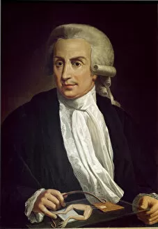
Background imageDissected Collection: Luigi Galvani with frog specimen (oil on canvas)

Luigi Galvani with frog specimen (oil on canvas)
LRI3725846 Luigi Galvani with frog specimen (oil on canvas) by Italian School, (18th century); Universita di Bologna, Italy; (add.info)

Background imageDissected Collection: The Dissecting Room, ca. 1838. Creator: T. C. Wilson

The Dissecting Room, ca. 1838. Creator: T. C. Wilson
The Dissecting Room, ca. 1838

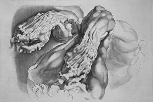
Background imageDissected Collection: Three Studies of Ecorches, after 1640. after 1640. Creator: Paulus Pontius

Three Studies of Ecorches, after 1640. after 1640. Creator: Paulus Pontius
Three Studies of Ecorches, after 1640

Background imageDissected Collection: Armpit muscles, 1831 artwork

Armpit muscles, 1831 artwork
Armpit muscles. Muscle connections in the second layer of the armpit. This anatomical artwork is plate 110 from volume 2 (1831) of Traite complet de l anatomie de l homme (1831-1854)

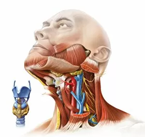
Background imageDissected Collection: Human neck anatomy, artwork C017 / 7259

Human neck anatomy, artwork C017 / 7259
Human neck anatomy, artwork. This dissection of the neck shows the larynx (voicebox) at the upper end of the trachea (windpipe, blue)

Background imageDissected Collection: Back Of The Male Human Body Showing Muscles Sinews And Bones From The Vessels Of The Human Body

Back Of The Male Human Body Showing Muscles Sinews And Bones From The Vessels Of The Human Body Edited By Jones Quain And William Wilson Published London By Taylor And Watson 1837

Background imageDissected Collection: Head And Shoulders Of The Male Human Body Showing Muscles Sinews And Bones From The Vessels Of The

Head And Shoulders Of The Male Human Body Showing Muscles Sinews And Bones From The Vessels Of The Human Body Edited By Jones Quain And William Wilson Published London By Taylor And Watson 1837

Background imageDissected Collection: Face and neck nerves, 1825 artwork

Face and neck nerves, 1825 artwork
Face and neck nerves. Head and neck dissected to show the superficial nerves (white). The parotid gland (normally present below the ear)

Background imageDissected Collection: Artery damage, MRA scan

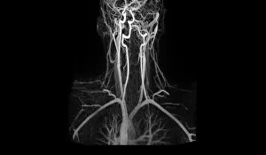
Artery damage, MRA scan
Artery damage. Magnetic resonance angiography (MRA) scan of the neck of a 23-year-old male motorcycle crash victim. Dissection of the right internal carotid artery (left)

Background imageDissected Collection: Brain meninges

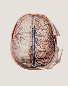
Brain meninges, historical anatomical artwork. This cranial (top) view of the brain (with front at the top) and its surrounding protective meninges, which have been partially dissected away

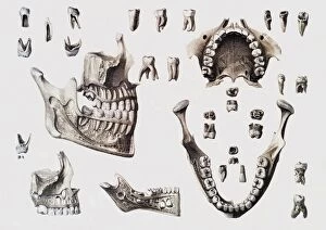
Background imageDissected Collection: Dental anatomy

Dental anatomy. Historical anatomical artwork of healthy and diseased human teeth and jaws. The teeth and jaws are seen from several different directions

Background imageDissected Collection: Blood vessels of chest and neck

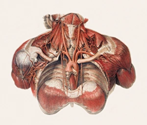
Blood vessels of chest and neck
Blood vessels of the chest and neck, historical anatomical artwork. The thoracic (chest) cavity has been dissected in this anterior (front) view to reveal part of the aorta

Background imageDissected Collection: Aortic dissection, 3D CT scan

Aortic dissection, 3D CT scan
Aortic dissection. 3D computed tomography (CT) scan of a 67-year-old man with an aortic dissection. The aorta, the bodys main artery, is running vertically down centre (red)

Background imageDissected Collection: Pigs heart

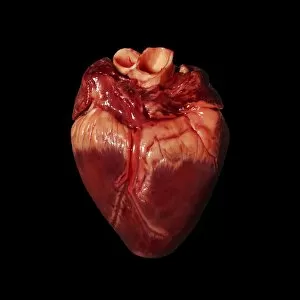
Pigs heart. The heart of a pig is anatomically similar to that of a human. The heart is a hollow muscular organ that pumps blood around the body

Background imageDissected Collection: Skeleton of a pigeon and a collection of pigeon feathers

Skeleton of a pigeon and a collection of pigeon feathers

Background imageDissected Collection: Cross section of a heart

Cross section of a heart
Cross section of a human heart showing the various chambers and valves. Date: 1910

Background imageDissected Collection: Cross-section through skeleton of Radiated tortoise (Astrochelys radiata), side view

Cross-section through skeleton of Radiated tortoise (Astrochelys radiata), side view

Background imageDissected Collection: Separated parts of human skull

Separated parts of human skull

Background imageDissected Collection: Twisted-pair cabling, side view

Twisted-pair cabling, side view

Background imageDissected Collection: Internal anatomy of frog, dissected with its internal organs hanging out of its belly

Internal anatomy of frog, dissected with its internal organs hanging out of its belly


All Professionally Made to Order for Quick Shipping

"Unveiling the Intricacies of Life: A Journey through Dissected Hints" Step into the world of Leonardo da Vinci's skull anatomy, where every curve and crevice holds secrets to our existence. Marvel at the intricate network of blood vessels that nourish our brain, as depicted in his mesmerizing artwork. The heart, a symbol of life itself, beats tirelessly within us. Explore its chambers and valves, understanding its vital role in sustaining our being. Witness the delicate dance between arteries and veins that carry life-giving oxygen throughout our bodies. Delve into history with an 1825 artwork showcasing male groin arteries - a testament to early anatomical studies. Discover how these hidden pathways contribute to our overall health and well-being. Venture further into the realm of dissection as we unravel the complex relationship between the heart and lungs. Witness their interdependence, understanding how they work harmoniously to ensure proper oxygenation for survival. Behold an 1825 artwork depicting head and chest arteries; a visual representation of scientific exploration from centuries past. Marvel at how these intricate networks sustain life by delivering essential nutrients throughout our bodies. Explore beyond mere surface appearances as we confront liver cirrhosis – a stark reminder of what can happen when this vital organ is compromised. Gain insight into medical conditions that challenge us while appreciating the resilience of human physiology. Witness scientists meticulously working with ragworm specimens – emblematic of tireless dedication towards unlocking nature's mysteries. Celebrate their unwavering commitment to advancing knowledge for generations to come. Embark on a journey through facial nerves, discovering their role in conveying emotions across countless expressions etched upon our faces. Appreciate how these invisible threads connect mind and body in profound ways. Transport yourself back in time to witness Eustachian tube syringing practices from the 18th century – an intriguing glimpse into historical medical techniques aimed at restoring balance within fragile auditory systems.