
Endocrinology Collection
"Exploring the Intricacies of Endocrinology: From Goitre to Hormones" In the realm of medical science


All Professionally Made to Order for Quick Shipping
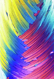
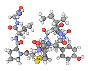
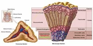
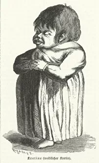
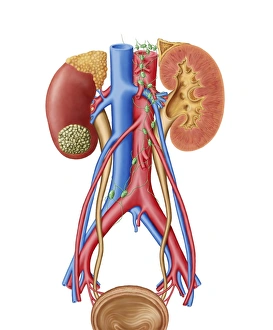
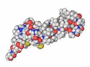
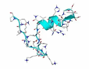
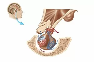
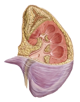
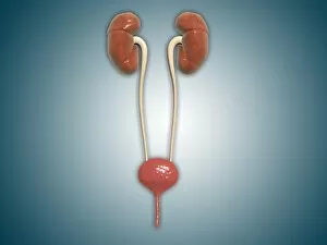
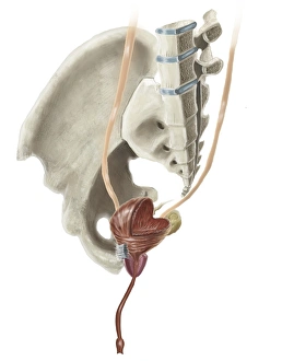
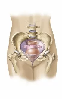
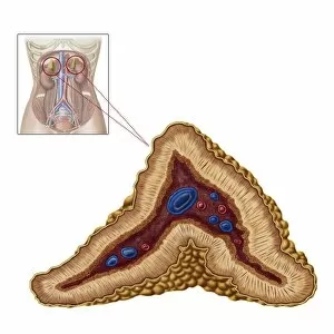
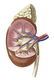
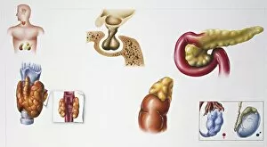
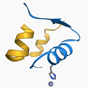
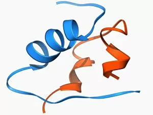
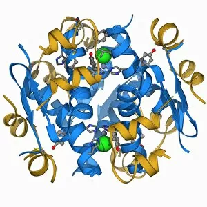
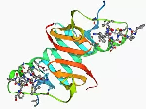
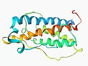
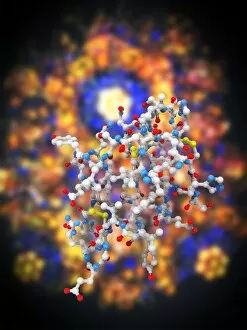
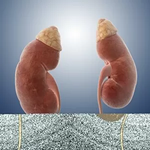
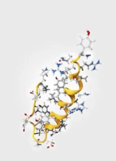
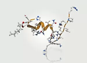
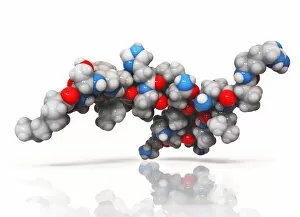
"Exploring the Intricacies of Endocrinology: From Goitre to Hormones" In the realm of medical science, endocrinology stands as a fascinating field that unravels the secrets behind our body's intricate hormonal system. Journeying back in time, we encounter a 15th-century artwork depicting a haunting goitre, shedding light on historical thyroid disorders. Delving deeper into this captivating subject, we explore cortisol crystals through a mesmerizing light micrograph. These tiny formations offer us a glimpse into the inner workings of our stress response and adrenal function. Moving forward, we stumble upon another intriguing light micrograph showcasing the delicate Islet of Langerhans. Nestled within our pancreas, these clusters of cells play an essential role in regulating blood sugar levels and are vital for managing diabetes. Shifting gears to neurotransmitters, we encounter oxytocin - often referred to as the "love hormone. " This molecule holds immense power over human bonding and social interactions, highlighting its significance in understanding emotional connections. Our exploration takes us further into thyroid anatomy with yet another captivating artwork. The intricate details depicted guide us through this crucial gland's structure and its impact on metabolism regulation. Returning to Islets of Langerhans but from an artistic perspective this time; their vibrant portrayal captures their importance in maintaining glucose balance within our bodies—a testament to both scientific beauty and functionality. Once again encountering cortisol crystals under microscopic lenses reveals more about stress-related conditions while reminding us how interconnected hormones are with our overall well-being. A poignant engraving portrays a woman suffering from congenital iodine deficiency syndrome or cretinism due to inadequate iodine intake during pregnancy—an alarming reminder of how nutrition plays an integral role in endocrine health. Crossing paths with an anatomical cross-section image showcases the complexity of adrenal glands—tiny powerhouses responsible for producing various hormones that regulate numerous bodily functions such as metabolism and stress response.