
Endometrium Collection
The endometrium, also known as the uterine lining, is a fascinating and crucial part of the female reproductive system


All Professionally Made to Order for Quick Shipping
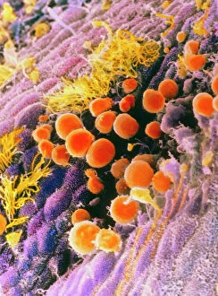
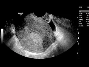
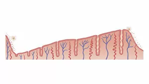
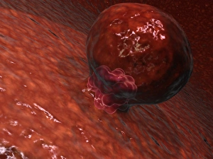
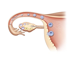
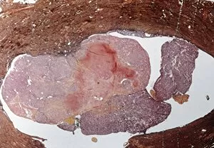
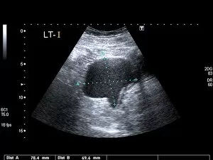
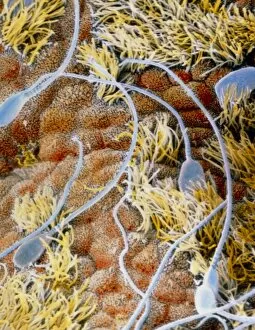
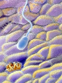
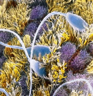
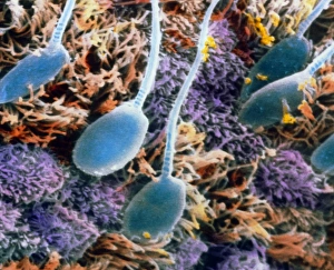
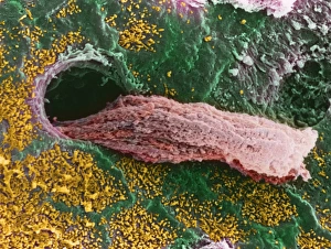
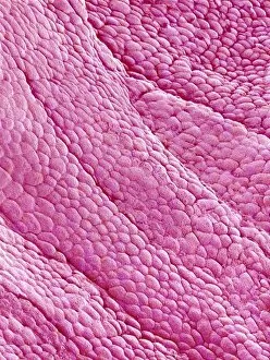
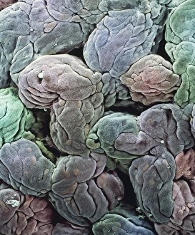
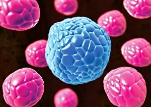
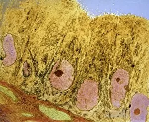
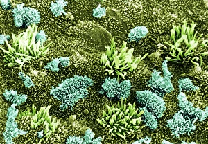
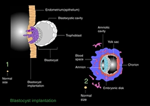
The endometrium, also known as the uterine lining, is a fascinating and crucial part of the female reproductive system. This SEM (scanning electron microscope) image showcases its intricate structure and vibrant colors, resembling a beautiful work of art. In this captivating internal cross-section of a human baby in the womb, we can see how the endometrium plays a vital role in supporting fetal development. It provides nourishment and protection to the growing embryo, ensuring its healthy growth. Biomedical illustrations like this one demonstrate an endometrial biopsy using a Novak curette. This procedure helps diagnose various conditions by examining tissue samples from the uterine lining. It allows doctors to detect abnormalities or potential issues that may require further investigation or treatment. During each menstrual cycle, the endometrium undergoes thickening in preparation for possible pregnancy. This biomedical illustration vividly depicts this process, showcasing how it transforms into a nurturing environment for implantation if fertilization occurs. Speaking of implantation, here we witness an incredible moment when a blastocyst begins attaching itself to the wall of the uterus. This pivotal event marks the beginning of pregnancy and highlights how essential a healthy endometrium is for successful conception. Unfortunately, sometimes complications arise within this delicate lining. Endometrial polyps are abnormal growths that can occur within it; X-rays C018/0587 and C018/0588 provide valuable insight into their diagnosis and management. Another condition affecting women's reproductive health is endometriosis - where tissue similar to that found in the uterus grows outside it. These X-rays (C014/4917 & C014/4916) help visualize these abnormal growths and aid medical professionals in formulating appropriate treatment plans. Ovulation and fertilization are key steps on every woman's journey towards motherhood. Through stunning artwork depicting these processes, we gain appreciation for how harmoniously orchestrated they must be for successful conception to occur.