Fraud Blocker Skip to main content

Enzyme Collection (page 8)

Enzymes: The Molecular Architects of Life DNA transcription, molecular model: they are the key players in the intricate process of DNA transcription

Background imageEnzyme Collection: Calcium ATPase ion pump C015 / 7143

Calcium ATPase ion pump C015 / 7143
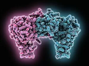
Calcium ATPase ion pump, molecular model. This enzyme is found in muscle cell membranes, where it pumps calcium in and out of muscle cells and controls muscle contractions

Background imageEnzyme Collection: Amylase, molecular model C015 / 7141

Amylase, molecular model C015 / 7141
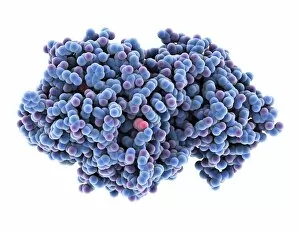
Amylase. Molecular model of the enzyme alpha-amylase from human saliva. Amylase catalyses the breakdown of starch to sugars, and so starts the digestive process

Background imageEnzyme Collection: Trypsin molecule with inhibitor C015 / 8436

Trypsin molecule with inhibitor C015 / 8436
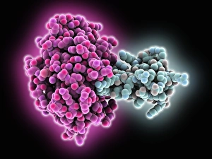
Trypsin molecule. Molecular model of the digestive protease enzyme beta-trypsin (pink) complexed with an inhibitor (blue)

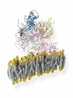
Background imageEnzyme Collection: Sodium-potassium ion pump proteins C015 / 9993

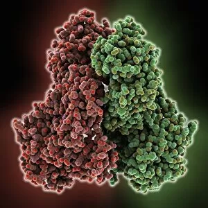
Sodium-potassium ion pump proteins C015 / 9993
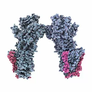
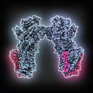
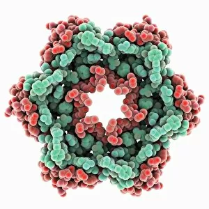
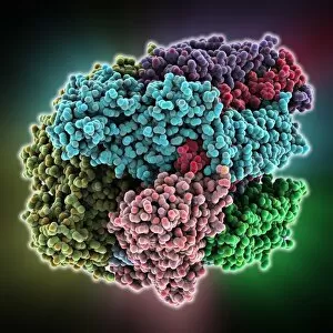
Sodium-potassium ion pump proteins, molecular model. Sodium-potassium ATPase (adenosine triphosphatase) is an ATP-powered ion pump found in all animal cells

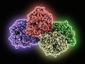
Background imageEnzyme Collection: Thrombin complexed with fibrinogen C015 / 7148

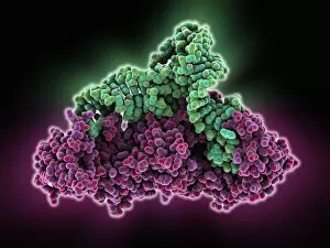
Thrombin complexed with fibrinogen C015 / 7148
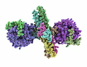
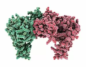
Thrombin complexed with fibrinogen, molecular model. The thrombin molecules (left and right, purple and green) are bound to the central part of the fibrinogen molecule (centre, multiple colours)

Background imageEnzyme Collection: Amylase, molecular model C015 / 7140

Amylase, molecular model C015 / 7140
Amylase. Molecular model of the enzyme alpha-amylase from human saliva. Amylase catalyses the breakdown of starch to sugars, and so starts the digestive process

Background imageEnzyme Collection: 3-hydroxyacyl-CoA dehydrogenase C015 / 9940

3-hydroxyacyl-CoA dehydrogenase C015 / 9940
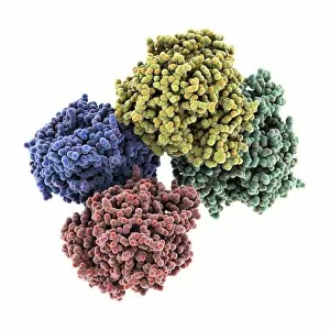
3-hydroxyacyl-CoA dehydrogenase, molecular model. This enzyme is found in human heart tissue, and catalyzes a reaction that is part of the beta-oxidation pathway

Background imageEnzyme Collection: Sodium-potassium ion pump proteins C015 / 9997

Sodium-potassium ion pump proteins C015 / 9997
Sodium-potassium ion pump proteins, molecular model. Sodium-potassium ATPase (adenosine triphosphatase) is an ATP-powered ion pump found in all animal cells

Background imageEnzyme Collection: Bacterial alcohol dehydrogenase molecule C015 / 7146

Bacterial alcohol dehydrogenase molecule C015 / 7146
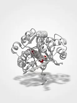
Bacterial alcohol dehydrogenase. Molecular model of an NADP-dependent alcohol dehydrogenase enzyme from the bacterium Thermoanaerobacter brockii

Background imageEnzyme Collection: Retinal-producing oxygenase enzyme C015 / 7150

Retinal-producing oxygenase enzyme C015 / 7150
Retinal-producing oxygenase enzyme, molecular model. This enzyme, an oxygenase, plays a role in the production and metabolism of retinal and other apocarotenoids

Background imageEnzyme Collection: Chymotrypsin inhibitor 2 molecule C015 / 8937

Chymotrypsin inhibitor 2 molecule C015 / 8937
Chymotrypsin inhibitor 2, molecular model. This enzyme is a serine protease inhibitor (serpin) found in barley (Hordeum vulgare) seeds

Background imageEnzyme Collection: Chymotrypsin inhibitor 2 molecule C015 / 8902

Chymotrypsin inhibitor 2 molecule C015 / 8902
Chymotrypsin inhibitor 2, molecular model. This enzyme is a serine protease inhibitor (serpin) found in barley (Hordeum vulgare) seeds

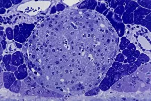
Background imageEnzyme Collection: Islet of Langerhans, light micrograph C016 / 0514

Islet of Langerhans, light micrograph C016 / 0514
Islet of Langerhans. Light micrograph of a section through an islet of Langerhans in a pancreas. Scattered throughout the pancreas, the islets are endocrine cells that secrete a range of hormones

Background imageEnzyme Collection: Thrombin complexed with fibrinogen C015 / 7149

Thrombin complexed with fibrinogen C015 / 7149
Thrombin complexed with fibrinogen, molecular model. The thrombin molecules (left and right, brown and pink) are bound to the central part of the fibrinogen molecule (centre, multiple colours)

Background imageEnzyme Collection: 3-hydroxyacyl-CoA dehydrogenase C015 / 9941

3-hydroxyacyl-CoA dehydrogenase C015 / 9941
3-hydroxyacyl-CoA dehydrogenase, molecular model. This enzyme is found in human heart tissue, and catalyzes a reaction that is part of the beta-oxidation pathway

Background imageEnzyme Collection: Calcium ATPase ion pump C015 / 7142

Calcium ATPase ion pump C015 / 7142
Calcium ATPase ion pump, molecular model. This enzyme is found in muscle cell membranes, where it pumps calcium in and out of muscle cells and controls muscle contractions

Background imageEnzyme Collection: Islet of Langerhans, light micrograph C016 / 0513

Islet of Langerhans, light micrograph C016 / 0513
Islet of Langerhans. Light micrograph of a section through an islet of Langerhans in a pancreas. Scattered throughout the pancreas, the islets are endocrine cells that secrete a range of hormones

Background imageEnzyme Collection: Trypsin molecule with inhibitor C015 / 8379

Trypsin molecule with inhibitor C015 / 8379
Trypsin molecule. Molecular model of the digestive protease enzyme beta-trypsin (pink) complexed with an inhibitor (blue)

Background imageEnzyme Collection: Retinal-producing oxygenase enzyme C015 / 7151

Retinal-producing oxygenase enzyme C015 / 7151
Retinal-producing oxygenase enzyme, molecular model. This enzyme, an oxygenase, plays a role in the production and metabolism of retinal and other apocarotenoids

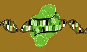
Background imageEnzyme Collection: Restriction enzyme and DNA, illustration C018 / 0785

Restriction enzyme and DNA, illustration C018 / 0785
Restriction enzyme. Illustration of a restriction enzyme (green) complexed with DNA (deoxyribonucleic acid, across centre)

Background imageEnzyme Collection: DNA repair, illustration C018 / 0782

DNA repair, illustration C018 / 0782
DNA repair. Illustation of a DNA (deoxyribonucleic acid) ligase enzyme (upper centre) repairing damaged DNA (spiral)

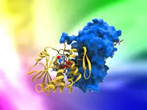
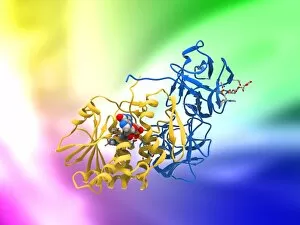
Background imageEnzyme Collection: Ricin molecule, artwork C017 / 3656

Ricin molecule, artwork C017 / 3656
Ricin molecule. Computer artwork showing the structure of a molecule of the toxic protein ricin. Ricin comprises two entwined amino acid chains; A (yellow) and B (blue)

Background imageEnzyme Collection: Ricin molecule, artwork C017 / 3655

Ricin molecule, artwork C017 / 3655
Ricin molecule. Computer artwork showing the structure of a molecule of the toxic protein ricin. Ricin comprises two entwined amino acid chains; A (yellow) and B (blue)

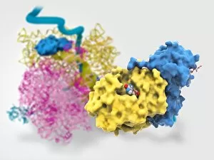
Background imageEnzyme Collection: Ricin molecule, artwork C017 / 3648

Ricin molecule, artwork C017 / 3648
Ricin molecule Computer artwork showing the structure of a molecule of the toxic protein ricin (blue and yellow) with an active ribosome in the background

Background imageEnzyme Collection: Aspartyl-tRNA synthetase protein molecule C014 / 0874

Aspartyl-tRNA synthetase protein molecule C014 / 0874
Aspartyl-tRNA synthetase protein molecule. Molecular model showing the structure of the active site of aspartyl-tRNA synthetase (DARS) from yeast

Background imageEnzyme Collection: Coagulation factor complex molecule C014 / 0409

Coagulation factor complex molecule C014 / 0409
Coagulation factor complex molecule. Molecular model showing the interaction between coagulation factor VIII (FVIII, pink, blue and yellow), factor IXa (FIXa)

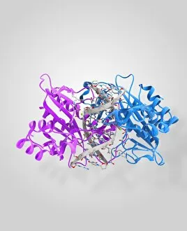
Background imageEnzyme Collection: EcoRV restriction enzyme molecule C014 / 2113

EcoRV restriction enzyme molecule C014 / 2113
EcoRV restriction enzyme. Molecular model of the type II restriction enzyme EcoRV (pink and blue) bound to a cleaved section of DNA (deoxyribonucleic acid, white)

Background imageEnzyme Collection: DNA helicase molecule

DNA helicase molecule
DNA helicase. Molecular model of a helicase molecule (blue) complexed with a molecule of DNA (deoxyribonucleic acid, pink and yellow)

Background imageEnzyme Collection: Methyladenine glycosylase bound to DNA C014 / 0877

Methyladenine glycosylase bound to DNA C014 / 0877
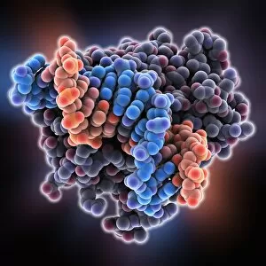
Methyladenine glycosylase bound to DNA. Computer model showing a molecule of human DNA-3-methyladenine glycosylase (purple) in complex with DNA (deoxyribonucleic acid, green and orange)

Background imageEnzyme Collection: EcoRV restriction enzyme molecule C014 / 2111

EcoRV restriction enzyme molecule C014 / 2111
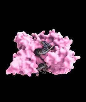
EcoRV restriction enzyme. Molecular model of the type II restriction enzyme EcoRV (pink) bound to a cleaved section of DNA (deoxyribonucleic acid, grey)

Background imageEnzyme Collection: Methyladenine glycosylase bound to DNA

Methyladenine glycosylase bound to DNA. Computer model showing a molecule of human DNA-3-methyladenine glycosylase (purple) in complex with DNA (deoxyribonucleic acid, blue and orange)

Background imageEnzyme Collection: EcoRV restriction enzyme molecule C014 / 2118

EcoRV restriction enzyme molecule C014 / 2118
EcoRV restriction enzyme. Molecular model of the type II restriction enzyme EcoRV (purple and beige) bound to a DNA molecule (deoxyribonucleic acid, yellow and orange)

Background imageEnzyme Collection: Coagulation factor complex molecule C014 / 0410

Coagulation factor complex molecule C014 / 0410
Coagulation factor complex molecule. Molecular model showing the interaction between coagulation factor VIII (FVIII, pink, blue and yellow), factor IXa (FIXa)

Background imageEnzyme Collection: Chymotrypsin inhibitor 2 molecule

Chymotrypsin inhibitor 2 molecule
Chymotrypsin inhibitor 2, molecular model. This plant enzyme is a serine protease inhibitor (serpin). It is thought that serpins in plants are a defence mechanism against insect and herbivore attack

Background imageEnzyme Collection: Stilbene synthase molecule C014 / 2292

Stilbene synthase molecule C014 / 2292
Stilbene synthase, molecular model. This enzyme is produced by some plants (including grapes, peanuts and blueberries) in response to stress from either ultraviolet light or certain fungi

Background imageEnzyme Collection: ATP synthase molecule

ATP synthase molecule. Molecular model showing the structure of ATP synthase (ATPase) subunit C. ATPase is an important enzyme that provides energy for cells through the synthesis of adenosine

Background imageEnzyme Collection: Stilbene synthase molecule C014 / 2291

Stilbene synthase molecule C014 / 2291
Stilbene synthase, molecular model. This enzyme is produced by some plants (including grapes, peanuts and blueberries) in response to stress from either ultraviolet light or certain fungi

Background imageEnzyme Collection: Saliva chemicals, molecular model

Saliva chemicals, molecular model
Saliva chemicals. Molecular structure of a group of saliva molecules. These include the antibody immunoglobulin A (blue, double-y shape, see C014/5652)

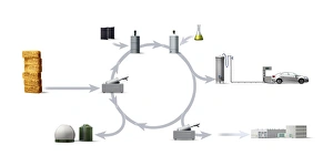
Background imageEnzyme Collection: Biofuel production and use, diagram

Biofuel production and use, diagram. At far left the first stage is the source material (biomass, plant or other organic material). This is added to a separation tank (centre left)

Background imageEnzyme Collection: Anthrax lethal factor molecule

Anthrax lethal factor molecule
Anthrax lethal factor, molecular model. This enzyme is one of three protein components that form the anthrax toxin produced by the bacterium Bacillus anthracis

Background imageEnzyme Collection: Alexander Fleming, caricature

Alexander Fleming, caricature
Alexander Fleming (1881-1955). Caricature of the Scottish biologist and pharmacologist Alexander Fleming holding a Petri dish

Background imageEnzyme Collection: Glutaminyl-tRNA synthetase molecule

Glutaminyl-tRNA synthetase molecule
Glutaminyl-tRNA synthetase protein molecule. Molecular model showing bacterial glutaminyl-tRNA synthetase complexed with glutamine tRNA (transfer ribonucleic acid)

Background imageEnzyme Collection: Kidney tissue, fluorescence micrograph C016 / 8484

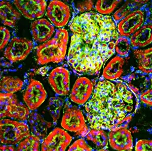
Kidney tissue, fluorescence micrograph C016 / 8484
Kidney tissue. Fluorescence deconvolution micrograph of a section through a kidney, showing glomeruli (green), cell nuclei (blue dots), and renal tubules (red, circular)

Background imageEnzyme Collection: Biotin ligase enzyme, molecular model

Biotin ligase enzyme, molecular model. This enzyme is a protein formed from 268 amino acids and two chains (green and pink)

Background imageEnzyme Collection: Methane monooxygenase enzyme

Methane monooxygenase enzyme, molecular model. This is the particulate methane monooxygenase (pMMO) form of this metalloenzyme, an integral membrane protein that contains copper and zinc

Background imageEnzyme Collection: FP2 malaria protease enzyme complex

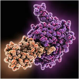
FP2 malaria protease enzyme complex, molecular model. This complex consists of the falcipain-2 (FP2) protease enzyme (purple, right) bound to a cystatin (orange, left), a form of protease inhibitor


All Professionally Made to Order for Quick Shipping

Enzymes: The Molecular Architects of Life DNA transcription, molecular model: they are the key players in the intricate process of DNA transcription, where they faithfully transcribe genetic information into RNA molecules. Metabolic enzyme, artwork: Metabolic enzymes act as catalysts in various biochemical reactions within our bodies, ensuring efficient metabolism and energy production. HIV reverse transcription enzyme: This remarkable enzyme allows the human immunodeficiency virus (HIV) to convert its RNA genome into DNA, enabling it to integrate with our own genetic material. Hepatitis C virus enzyme, molecular model: Understanding the structure and function of hepatitis C virus enzymes is crucial for developing effective treatments against this persistent viral infection. Anatomy of Organs Engraving: Enzymes play a vital role in maintaining organ health by facilitating essential processes like digestion, respiration, and hormone regulation throughout our body's intricate anatomy. Manganese superoxide dismutase enzyme F006 / 9423: This antioxidant enzyme protects cells from harmful free radicals by converting them into less damaging substances—a guardian against oxidative stress. Cytochrome b5 molecule C015 / 6696: As an electron carrier protein found in cell membranes, cytochrome b5 assists other enzymes in performing critical metabolic reactions involved in energy production and lipid metabolism. Glutamine synthetase enzyme: Essential for nitrogen metabolism, glutamine synthetase ensures that ammonia produced during cellular processes is safely converted into non-toxic compounds like amino acids or urea. RNA-editing enzyme, molecular model: These specialized enzymes modify RNA molecules after their synthesis—fine-tuning gene expression patterns and expanding the diversity of proteins encoded by our genes. ATPase molecule: ATPases are indispensable for cellular energy transfer; these enzymes hydrolyze adenosine triphosphate (ATP), releasing stored energy to power various cellular processes.