
Epidermal Collection
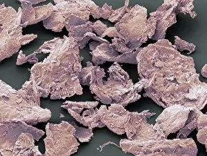
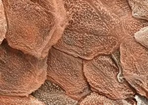
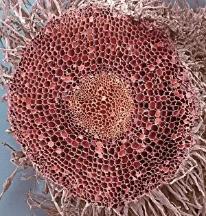
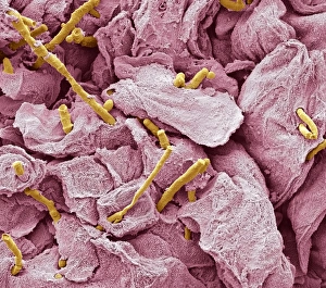
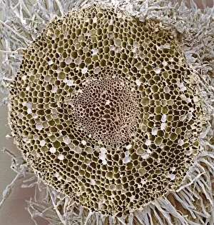
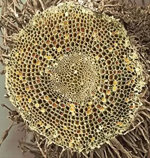
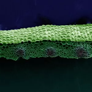
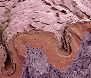
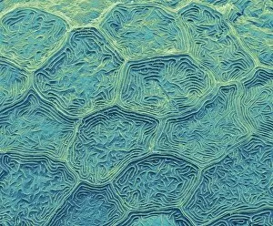
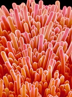
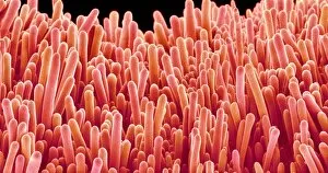
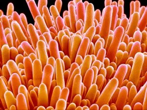
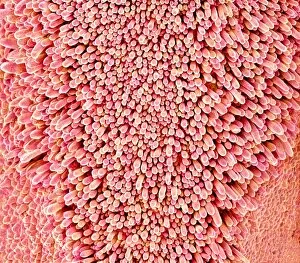
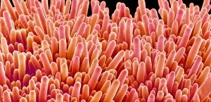
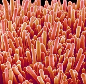
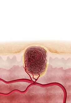
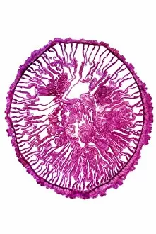
"Exploring the Intricate World Surfaces: From Periwinkle Petals to Zebra Fish Skin" The epidermis, or outermost layer of our skin and various plant surfaces


All Professionally Made to Order for Quick Shipping
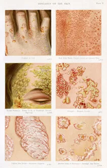
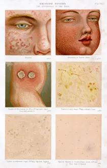
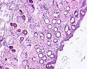
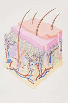
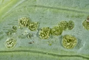
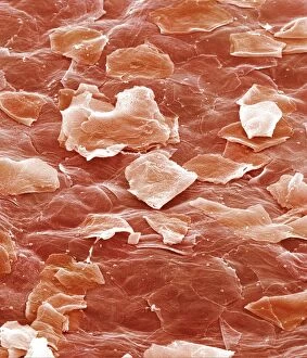
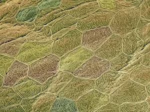
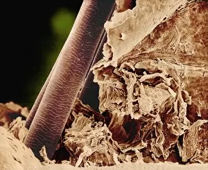
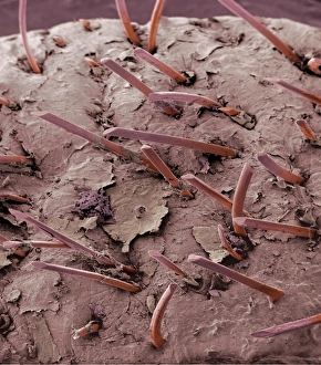
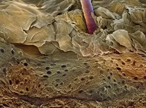
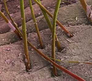
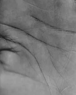
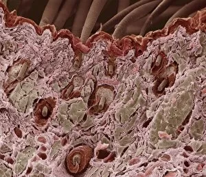
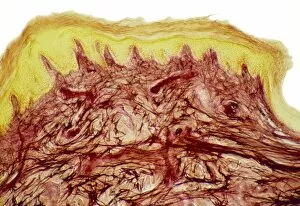
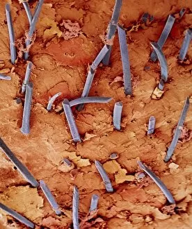
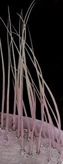
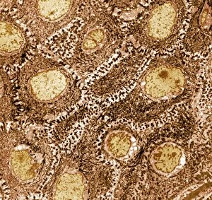
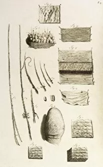
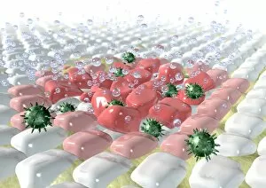
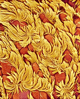
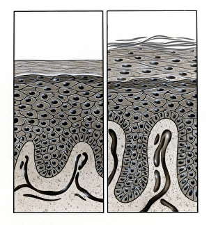
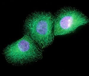
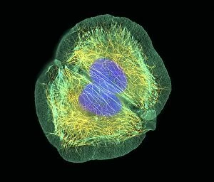
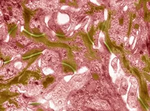
"Exploring the Intricate World Surfaces: From Periwinkle Petals to Zebra Fish Skin" The epidermis, or outermost layer of our skin and various plant surfaces, is a fascinating realm that holds secrets waiting to be discovered. Through powerful imaging techniques such as Scanning Electron Microscopy (SEM) and Transmission Electron Microscopy (TEM), we can delve into the intricate details of these surfaces. In one captivating image, the delicate periwinkle petal surface reveals its mesmerizing beauty under SEM. The magnified view showcases the exquisite patterns and textures that make each petal unique. Moving on to zebra fish skin, SEM unravels a world unseen by the naked eye. The scales resemble an artistic mosaic, forming a protective shield for these aquatic creatures. Zooming in further with TEM, we witness the astonishing complexity of skin cells. These microscopic building blocks play a vital role in maintaining our body's integrity while orchestrating essential functions like protection and sensation. Shifting gears from animals to plants again, an orchid petal captured under SEM unveils nature's artistry at its finest. Delicate structures interweave seamlessly, creating breathtaking displays of color and form. Next up are olive leaf trichomes observed through SEM. These tiny hair-like projections serve as armor against predators while also aiding in moisture retention for this resilient plant species. A pansy petal takes center stage once more under SEM's watchful lens. Its velvety texture comes alive as we explore every nook and cranny meticulously designed by Mother Nature herself. Venturing beyond natural beauty lies medical significance - Diseases of the Skin plates 4 & 5 showcase dermatological conditions that affect human epidermis. Eruptive Fevers plate 6 provides insight into infectious diseases impacting our largest organ –the skin– highlighting their symptoms for better understanding and diagnosis.