
Epidermis Collection
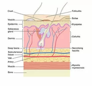
The epidermis, our body's protective outer layer, is a fascinating subject that encompasses various aspects of skin disorders and artwork


All Professionally Made to Order for Quick Shipping
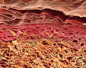
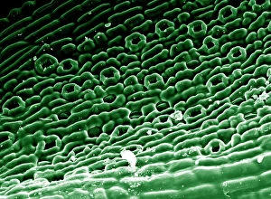
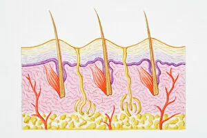
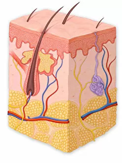
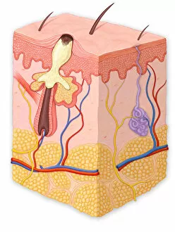
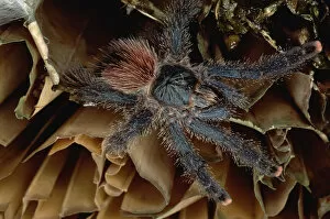
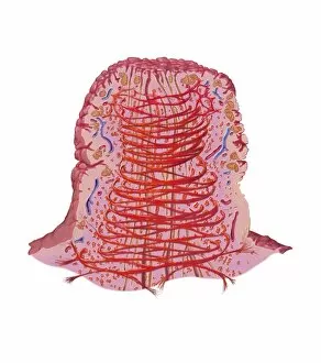
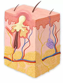
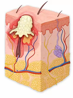
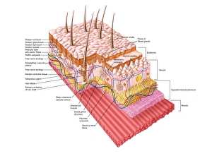
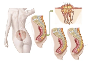
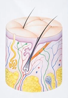
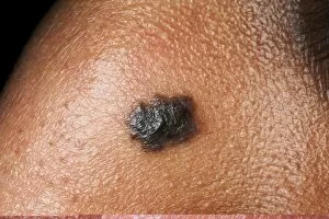
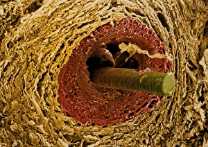
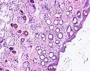
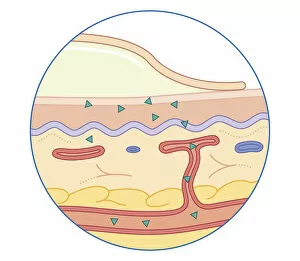
The epidermis, our body's protective outer layer, is a fascinating subject that encompasses various aspects of skin disorders and artwork. From examining the lime tree stem under a light micrograph to exploring a section through human skin using scanning electron microscopy (SEM), there are endless possibilities for understanding this intricate organ. In Picture No. 11675585, we can observe an illustration depicting the cross-section of human skin with heat trapped by erect hairs. This visual representation highlights the complexity of our epidermis and its role in regulating body temperature. Delving deeper into the layers of our skin, SEM images reveal the intricate patterns and structures present on its surface. Whether it's studying the texture of tomato leaf or periwinkle petal surfaces or even analyzing zebra fish skin at a microscopic level, these images provide valuable insights into how diverse organisms have adapted their epidermal layers for survival. Examining individual cells within the epidermis becomes possible through transmission electron microscopy (TEM). By zooming in on these tiny building blocks, we gain insight into their structure and function, helping us understand how they contribute to overall skin health. Even beyond humans, other organisms like olive leaves also possess unique features worth exploring. Through SEM imaging of olive leaf trichomes - small hair-like structures covering their surface - we can appreciate nature's intricacies while drawing parallels between different species' adaptations. From understanding common skin disorders to appreciating art inspired by this remarkable organ's beauty and functionality, delving into the world opens up a realm filled with scientific wonder and artistic inspiration alike.