
Erythrocyte Collection
"Erythrocyte: The Lifeline of Blood" Erythrocytes, commonly known as red blood cells, play a crucial role in our circulatory system


All Professionally Made to Order for Quick Shipping
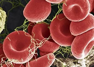
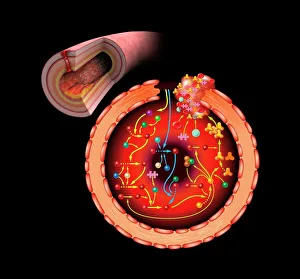
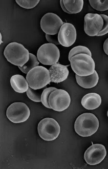
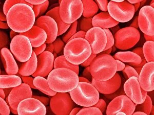
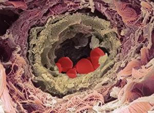
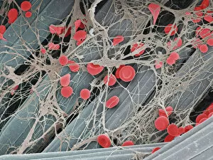
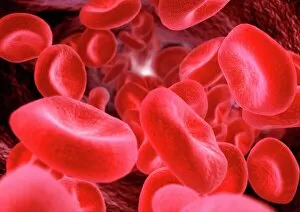
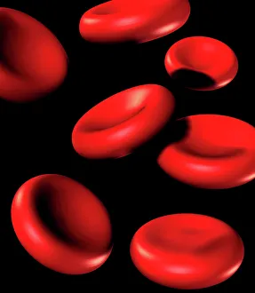
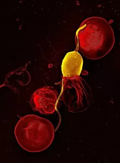
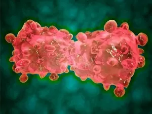
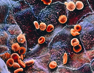
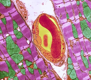
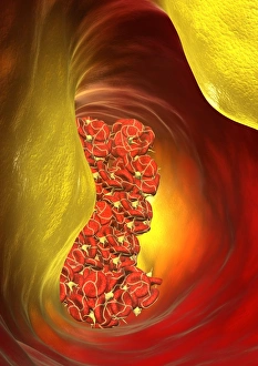
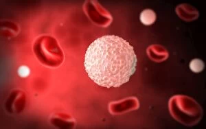
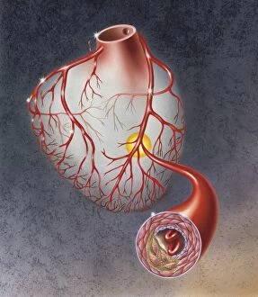
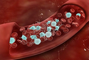
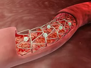
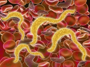
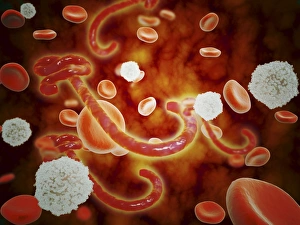
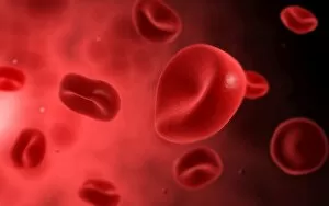
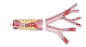
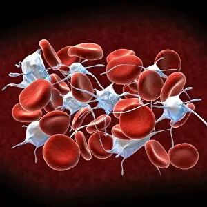
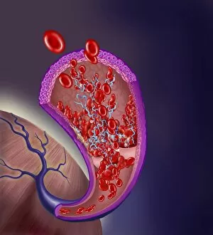
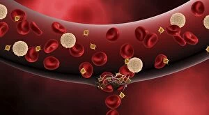
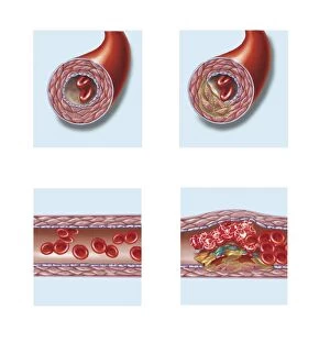
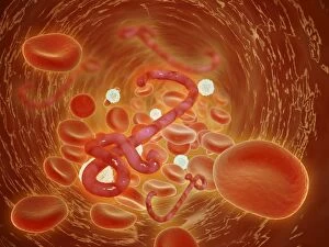
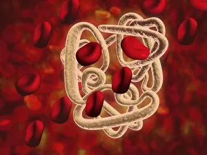
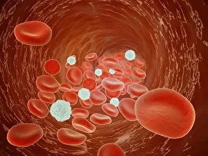
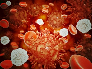
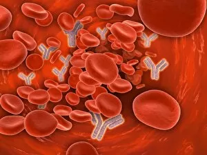
"Erythrocyte: The Lifeline of Blood" Erythrocytes, commonly known as red blood cells, play a crucial role in our circulatory system. These tiny, disc-shaped cells are responsible for transporting oxygen from the lungs to every tissue and organ in our body. During menstruation, the uterus lining sheds and erythrocytes come into action. They help deliver necessary nutrients and oxygen to support the regrowth of this lining, ensuring a healthy reproductive cycle. Under scanning electron microscopy (SEM), these remarkable blood cells reveal their intricate structure. Their biconcave shape allows for maximum surface area exposure, facilitating efficient gas exchange within our bodies. The importance of erythrocytes extends beyond just oxygen transport. They also contribute to the complex process of blood coagulation cascade – an essential mechanism that prevents excessive bleeding when injuries occur. Artwork C016/9873 beautifully illustrates this intricate cascade that leads to clot formation. In SEM image C016/9747, we witness a close-up view of a blood clot formed by platelets and fibrin strands working together harmoniously to seal wounds effectively. Human red blood corpuscles captured under SEM showcase their vibrant nature while reminding us of their vital function within our bloodstream - maintaining homeostasis by carrying carbon dioxide back to the lungs for elimination. Even parasites like mouse malaria can be observed through SEM attached to erythrocytes - highlighting how these cells serve as hosts during infection but also become targets for immune responses against such invaders. Blood vessels act as highways where erythrocytes travel tirelessly throughout our bodies. These microscopic highways ensure proper distribution of nutrients and removal of waste products from various tissues they reach. SEM images further unveil intriguing details about erythrocyte behavior; one such example is seen in an image depicting a blood clot forming on plaster's surface – showcasing how these incredible cells respond swiftly when faced with injury or damage outside the body too.