Fraud Blocker Skip to main content

False Coloured Collection (page 3)

"Revealing the Unseen: Exploring the World Through False Colors" Step back in time to medieval alchemy

Background imageFalse Coloured Collection: Red blood cells, SEM

Red blood cells, SEM
Red blood cells, coloured scanning electron micrograph (SEM). Red blood cells (erythrocytes) are biconcave, giving them a large surface area for gas exchange, and highly elastic

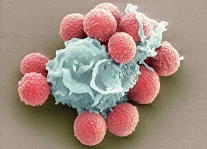
Background imageFalse Coloured Collection: Phagocytosis of fungal spores, SEM

Phagocytosis of fungal spores, SEM
Phagocytosis of a fungus spore. Coloured scanning electron micrograph (SEM) of a neutrophil (blue) phagocytosing (engulfing and destroying) spores from the fungus Aspergillus fumigatus (red)

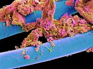
Background imageFalse Coloured Collection: Used dental floss, SEM

Used dental floss, SEM
Used dental floss. Coloured scanning electron micrograph (SEM) of dental plaque on strands of used dental floss. Plaque consists of a film of bacteria embedded in a glycoprotein matrix

Background imageFalse Coloured Collection: Fractured jawbone, X-ray

Fractured jawbone, X-ray
Fractured jawbone. Coloured X-ray of a human jaw showing a fractured lower jawbone (mandible). The fracture has occurred to the left side of the lower jawbone (bottom right, orange)

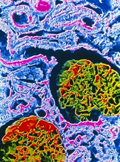
Background imageFalse Coloured Collection: False-colour SEM showing two renal glomeruli

False-colour SEM showing two renal glomeruli
False-colour scanning electron micrograph of a section of kidney cortex, showing two glomeruli (red & green). A glomerulus is a tiny knot of blood capillaries which lies cupped within a Bowmans

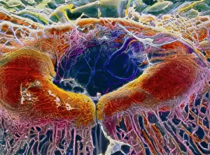
Background imageFalse Coloured Collection: False-colour SEM of retina featuring central fovea

False-colour SEM of retina featuring central fovea
Foveal retina. False-colour scanning electron micrograph of the human retina featuring the central fovea, a crater-like depression in the photosensitive layer of the eye

Background imageFalse Coloured Collection: Myelination of nerve fibres, TEM

Myelination of nerve fibres, TEM
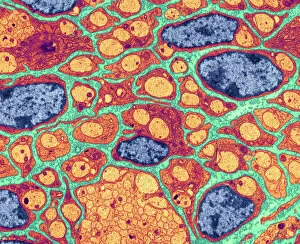
Myelination of nerve fibres. Coloured transmission electron micrograph (TEM) of Schwann cells (red, with blue nuclei) insulating nerve fibres (axons, orange) with a myelin sheath

Background imageFalse Coloured Collection: Myelinated nerve, TEM

Myelinated nerve, TEM
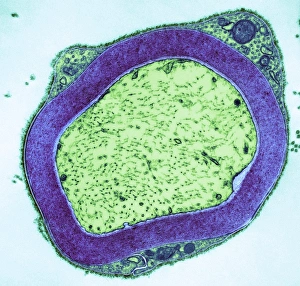
Myelinated nerve. Coloured transmission electron micrograph (TEM) of myelinated nerve fibres and Schwann cells. Myelin (purple) is an insulating fatty layer that surrounds nerve fibres (axons)

Background imageFalse Coloured Collection: Neutrophil cell trapping bacteria, SEM

Neutrophil cell trapping bacteria, SEM
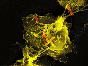
Neutrophil cell trapping bacteria. Coloured scanning electron micrograph (SEM) of bacteria (rod-shaped) being trapped by a neutrophil cell

Background imageFalse Coloured Collection: Royal Vauxhall pre-flight consultation

Royal Vauxhall pre-flight consultation. Historical artwork depicting a meeting held prior to the flight of the Royal Vauxhall in 1836

Background imageFalse Coloured Collection: Cauliflower head, SEM

Cauliflower head, SEM
Cauliflower. Coloured scanning electron micrograph (SEM) of the head of a cauliflower (Brassica oleracea botrytis). The arrangement of florets around the head is according to a Fibonacci series

Background imageFalse Coloured Collection: Mitochondrion, TEM

Mitochondrion, TEM
Mitochondrion. Coloured transmission electron micrograph (TEM) of mitochondria (green/blue) in heart muscle. Mitochondria are organelles found in the cytoplasm of eukaryotic cells

Background imageFalse Coloured Collection: Healthy brain, MRI scans

Healthy brain, MRI scans
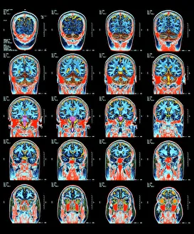
Healthy brain. Coloured magnetic resonance imaging (MRI) scans through the head of a 55 year old man, showing the brain sectioned in coronal (frontal) view

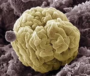
Background imageFalse Coloured Collection: Kidney glomerulus, SEM

Kidney glomerulus, SEM
Kidney glomerulus, coloured scanning electron micrograph (SEM). Podocyte cells, which make up the surface of the glomerulus, are yellow. Magnification: x650 when printed at 10 centimetres wide

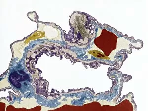
Background imageFalse Coloured Collection: Lung tissue, TEM

Lung tissue, TEM
Lung tissue, coloured transmission electron micrograph (TEM). A single alveolus is at center. At top right is a pulmonary capillary containing a red blood cell (red) and a platelet (yellow)

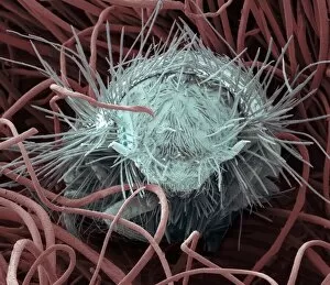
Background imageFalse Coloured Collection: Carpet beetle larva, SEM

Carpet beetle larva, SEM
Carpet beetle larva. Coloured scanning electron micrograph (SEM) of a carpet beetle (Anthrenus sp.) larva amongst the fibres of a wool carpet

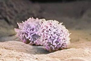
Background imageFalse Coloured Collection: Macrophage cells, SEM

Macrophage cells, SEM
Macrophage cells. Coloured scanning electron micrograph (SEM) of macrophage white blood cells. Macrophages are cells of the bodys immune system

Background imageFalse Coloured Collection: Influenza virus particles, TEM

Influenza virus particles, TEM
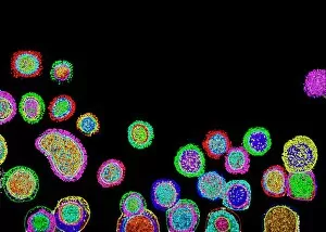
Influenza virus particles, coloured transmission electron micrograph (TEM). This virus is the cause of the respiratory disease influenza (flu) in humans and other animals

Background imageFalse Coloured Collection: Hepatitis C viruses, TEM

Hepatitis C viruses, TEM
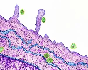
Hepatitis C viruses. Coloured transmission electron micrograph (TEM) of hepatitis C virus particles (green) infecting cultured liver cells (purple)

Background imageFalse Coloured Collection: Ovarian follicle, SEM

Ovarian follicle, SEM
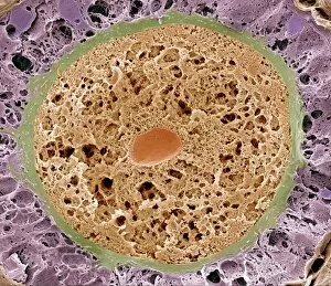
Ovarian follicle. Coloured scanning electron micrograph (SEM) of a fracture through a secondary follicle in the ovary. The oocyte (developing egg) is orange and its central nucleus is darker orange

Background imageFalse Coloured Collection: Dinoflagellate plankton, SEM

Dinoflagellate plankton, SEM
Dinoflagellate plankton. Coloured scanning electron micrograph (SEM) of the calcareous (calcium-based) test (shell) of a dinoflagellate Calciodinellum sp

Background imageFalse Coloured Collection: Spiny spider, SEM

Spiny spider, SEM
Spiny spider. Coloured scanning electron micrograph (SEM) of a spiny spider (Gasteracantha sp.), showing its characteristic spines (red) and large mouthparts (grey, centre left)

Background imageFalse Coloured Collection: Trachea lining, SEM

Trachea lining, SEM
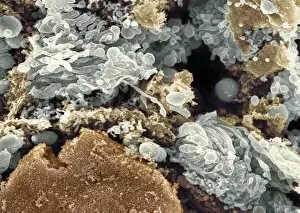
Trachea lining, coloured scanning electron micrograph (SEM). The tracheal lining contains mucus-secreting goblet cells and epithelial cells bearing hair-like cilia (blue)

Background imageFalse Coloured Collection: Dandelion pollen grain, SEM

Dandelion pollen grain, SEM
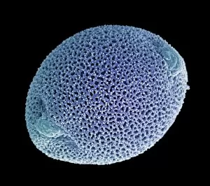
Dandelion (Taraxacum officinale) pollen grain, coloured scanning electron micrograph (SEM). Pollen grains are reproductive structures produced by the male parts of flowering plants

Background imageFalse Coloured Collection: Golgi apparatus, SEM

Golgi apparatus, SEM
Golgi apparatus, coloured scanning electron micrograph (SEM). Section through a liver cell showing its Golgi apparatus (grey), a membrane-bound organelle that modifies and packages proteins

Background imageFalse Coloured Collection: Goosegrass fruits, SEM

Goosegrass fruits, SEM
Goosegrass fruits. Coloured scanning electron micrograph (SEM) of the hooked fruits of a goosegrass plant (Galium aparine)

Background imageFalse Coloured Collection: Synapse nerve junctions, SEM

Synapse nerve junctions, SEM
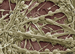
Synapse nerve junctions. Coloured scanning electron micrograph (SEM) of nerve cells showing the synapses (junctions, bulges) between them

Background imageFalse Coloured Collection: Silver birch twig, SEM

Silver birch twig, SEM
Silver birch twig. Coloured scanning electron micrograph (SEM) of a section through a silver birch (Betula pendula) twig. At the centre of the twig (top centre left) is the pith

Background imageFalse Coloured Collection: Pollen on a bee leg, SEM

Pollen on a bee leg, SEM
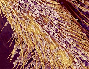
Pollen on a bee leg. Coloured scanning electron micrograph (SEM) of pollen grains (pale purple) on the leg of a bee (superfamily Apoidea). Pollen grains are the male sex cells of a flowering plant

Background imageFalse Coloured Collection: Pseudomonas aeruginosa bacteria, SEM

Pseudomonas aeruginosa bacteria, SEM
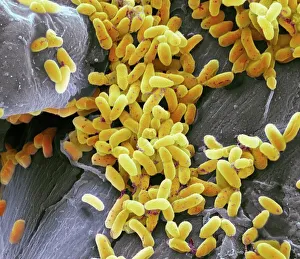
Pseudomonas aeruginosa bacteria, coloured scanning electron micrograph (SEM). These Gram-negative rod-shaped bacteria are found in soil and water, and as normal flora in the human intestine

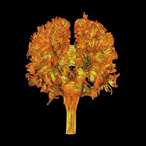
Background imageFalse Coloured Collection: White matter fibres, DTI scan

White matter fibres, DTI scan
White matter fibres. Coloured 3D diffusion tensor imaging (DTI) scan of the brain, showing the bundles of white matter nerve fibres

Background imageFalse Coloured Collection: Fly on sugar, SEM

Fly on sugar, SEM
Fly on sugar, coloured scanning electron micrograph (SEM). Flies (order Diptera) are insects that can spread disease when they feed on human food

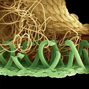
Background imageFalse Coloured Collection: SEM of a hooks and loops fastener

SEM of a hooks and loops fastener
Coloured scanning electron micrograph (SEM)of a hooks and loops fastener. It is a two-sheet material that is used as a reversible fastener on clothing and fabrics

Background imageFalse Coloured Collection: Magnolia (Magnolia virginiana) flower head, X-ray

Magnolia (Magnolia virginiana) flower head, X-ray
Magnolia (Magnolia virginiana) flower head, coloured X-ray

Background imageFalse Coloured Collection: Great coneflower (Rudbeckia maxima), X-ray

Great coneflower (Rudbeckia maxima), X-ray
Great coneflower (Rudbeckia maxima), coloured X-ray

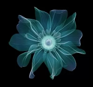
Background imageFalse Coloured Collection: Hellebore (Helleborus hybridis), X-ray

Hellebore (Helleborus hybridis), X-ray
Hellebore (Helleborus hybridis), coloured X-ray

Background imageFalse Coloured Collection: Dinoflagellates, SEM

Dinoflagellates, SEM
Dinoflagellates. Coloured scanning electron micrograph (SEM) of Prorocentrum dinoflagellates (round) and Ceratium dinoflagellates (branched). Dinoflagellates are unicellular protozoans

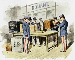
Background imageFalse Coloured Collection: X-ray security machine, 1900

X-ray security machine, 1900
X-ray security machine. Historical artwork of a fluoroscope being used in a late 19th-century French customs hall. The fluoroscope was designed and built by the French inventor Eugene Ducretet

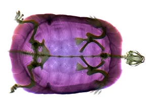
Background imageFalse Coloured Collection: Turtle

Turtle, coloured X-ray. The head of the turtle is at right
Background imageFalse Coloured Collection: Red blood cells, SEM

Red blood cells, SEM
Red blood cells. Coloured scanning electron micrograph (SEM) of red blood cells (erythrocytes). Red blood cells are biconcave, disc-shaped cells that transport oxygen from the lungs to body cells
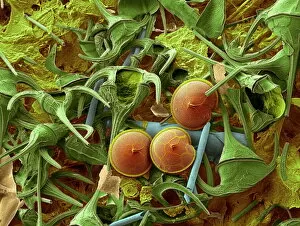
Background imageFalse Coloured Collection: Diatom frustule, SEM

Diatom frustule, SEM
Diatom frustule. Coloured scanning electron micrograph (SEM) of the frustule of a Pinnularia sp. diatom. The diatoms are a group of photosynthetic, single-celled algae containing about 10

Background imageFalse Coloured Collection: Pine tree stem, light micrograph

Pine tree stem, light micrograph
Pine tree stem. Coloured light micrograph of a cross-section through the stem of a pine tree. (Pinus sp.). The red wood (xylem, lower left) has resin canals in it, which are outlined in green

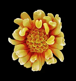
Background imageFalse Coloured Collection: Buttercup flower, SEM

Buttercup flower, SEM
Buttercup flower (Ranunculus sp.), coloured scanning electron micrograph (SEM). The pistil (centre) is surrounded by the stamens (yellow)

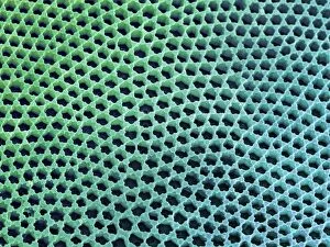
Background imageFalse Coloured Collection: Diatom cell wall, SEM

Diatom cell wall, SEM
Diatom cell wall. Coloured scanning electron micrograph (SEM) of the pattern of small holes, called striae, in the cell wall of a diatom alga. This is a Coscinodiscus sp. diatom

Background imageFalse Coloured Collection: Pharos lighthouse, Ptolemaic Egypt

Pharos lighthouse, Ptolemaic Egypt
Pharos lighthouse. Historical artwork showing what the Pharos lighthouse at Alexandria, Egypt, in the third century BC, might have looked like

Background imageFalse Coloured Collection: Nose and sinuses, CT scan

Nose and sinuses, CT scan
Nose anatomy and sinuses. Coloured computed tomography (CT) scan of a frontal view through the nose of a 31 year old woman

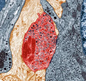
Background imageFalse Coloured Collection: Nerve synapse, TEM

Nerve synapse, TEM
Nerve synapse. Coloured transmission electron micrograph (TEM) of the neuron (nerve) terminal at a synapse in the diaphragm

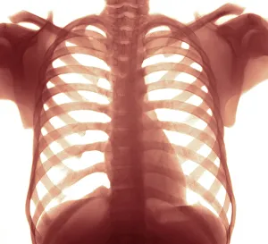
Background imageFalse Coloured Collection: Chest X-ray of a healhty human heart

Chest X-ray of a healhty human heart
Chest X-ray. Coloured X-ray of a healthy human heart. The heart is the dark area to the lower right of the spinal column. Chest X-rays can be used to diagnose diseases of the heart


All Professionally Made to Order for Quick Shipping

"Revealing the Unseen: Exploring the World Through False Colors" Step back in time to medieval alchemy, where a peculiar contraption known as the "Medieval urine wheel" was used to extract hidden secrets from bodily fluids. Fast forward to modern times and witness how science has evolved, unveiling breathtaking wonders like the Orion Nebula. Delve into the microscopic realm, where particle tracks paint a mesmerizing picture of subatomic interactions. Journey across Martian landscapes through the eyes of Spirit rover images, capturing otherworldly vistas that ignite our imagination. Peering into our own genetic blueprint, false colors bring clarity to X and Y chromosomes, unraveling mysteries within our very cells. A horse's skull takes on an ethereal glow as we explore its intricate structure using advanced imaging techniques. Witness synapse nerve junctions come alive with vibrant hues under a transmission electron microscope (TEM), revealing connections that shape our thoughts and actions. A person holding a camera becomes an enigma when captured through X-ray technology – their inner world exposed for all to see. Marvel at tardigrades' resilience as they navigate their microscopic universe under scanning electron microscopy (SEM). Calcareous phytoplankton dance with color in intricate patterns while nerve cells reveal their complexity in stunning detail. Travel back in time to 19th-century Cornwall and discover the harsh reality of tin mining brought vividly to life through false colors – reminding us of humanity's enduring spirit even amidst adversity. In this captivating journey through various realms – from ancient alchemy to distant galaxies, minuscule particles to majestic landscapes – false colors unlock hidden dimensions beyond what meets the eye, inviting us all into a world brimming with wonder and discovery.