Fraud Blocker Skip to main content

Fibula Collection

The fibula, a slender bone in the human leg, plays a vital role in supporting the anatomy of the knee joint

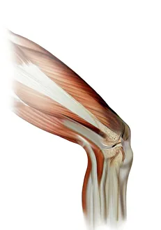
Background imageFibula Collection: Anatomy of human knee joint

Anatomy of human knee joint

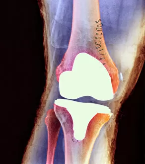
Background imageFibula Collection: Knee joint prosthesis, X-ray

Knee joint prosthesis, X-ray
Knee joint replacement. Coloured X-ray of the knee of a 56 year old man (front view) showing an artificial (prosthetic) joint replacing the knee joint

Background imageFibula Collection: Damaged knee ligament, artwork

Damaged knee ligament, artwork
Damaged knee ligament. Computer artwork of the muscle structure of the leg and anatomy of the human knee joint, showing a tear (dark line, centre right) in one of the cruciate ligaments (beige)

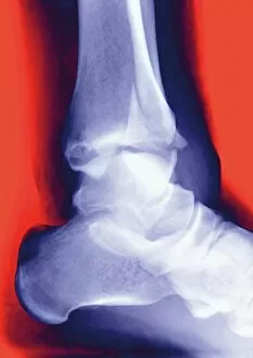
Background imageFibula Collection: Fractured ankle, X-ray

Fractured ankle, X-ray
Fractured ankle. Coloured profile X-ray of a distal fibula fracture (upper centre). The fibula is the smaller leg bone running down from top centre. The larger bone is the tibia

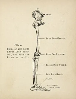
Background imageFibula Collection: Diagram of the bones of the right leg and hip

Diagram of the bones of the right leg and hip
Diagram of the bones of the right leg, showing the joint with the pelvis at the hip

Background imageFibula Collection: Healthy knee, X-ray

Healthy knee, X-ray
Healthy knee. Coloured frontal (right) and profile (left) X-rays of the healthy knee of a 24 year old patient

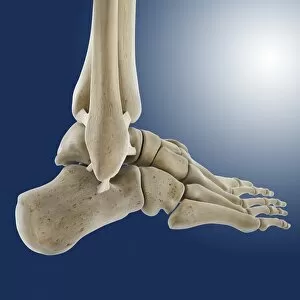
Background imageFibula Collection: Outer ankle ligaments, artwork C013 / 4452

Outer ankle ligaments, artwork C013 / 4452
Outer ankle ligaments. Computer artwork of the bones and ligaments (white) of the right foot and ankle seen from the side

Background imageFibula Collection: Knee joint

Knee joint with cruciate ligaments

Background imageFibula Collection: Inner ankle ligaments, artwork C013 / 4451

Inner ankle ligaments, artwork C013 / 4451
Inner ankle ligaments. Computer artwork of the bones and ligaments (white) of the right foot and ankle seen from the side

Background imageFibula Collection: Total knee replacement, X-rays

Total knee replacement, X-rays
Total knee replacement. Coloured frontal (left) and profile (right) X-rays of the right knee of a 69 year old patient after total knee replacement surgery

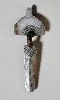
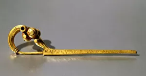
Background imageFibula Collection: Fibule d'arc, art wisigoth, 7th century

Fibule d'arc, art wisigoth, 7th century
CUL4852717 Fibule d'arc, art wisigoth, 7th century by Visigothic, (7th century); Archaeological Museum of Catalonia, Barcelona, Spain; (add.info.: Fibule d'arc, art wisigoth)

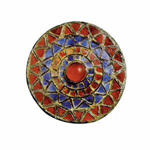
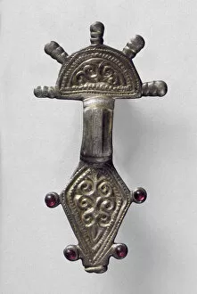
Background imageFibula Collection: Circular fibule dotted with glass, art visigoth, 7th century

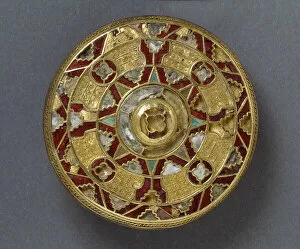
Circular fibule dotted with glass, art visigoth, 7th century
CUL4852685 Circular fibule dotted with glass, art visigoth, 7th century by Visigothic, (7th century); Archaeological Museum of Catalonia, Barcelona

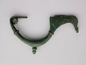
Background imageFibula Collection: Fibula with Architectural Design, Roman Period (30 BCE-395 CE) or later. Creator: Unknown

Fibula with Architectural Design, Roman Period (30 BCE-395 CE) or later. Creator: Unknown
Fibula with Architectural Design, Roman Period (30 BCE-395 CE) or later

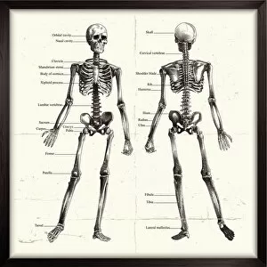
Background imageFibula Collection: Labelled Human Skeleton. Engraving

Labelled Human Skeleton. Engraving
Labelled engraved image of human skeleton

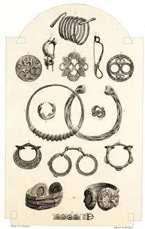
Background imageFibula Collection: Celtic Relics, Personal Ornaments of Gold and Bronze

Celtic Relics, Personal Ornaments of Gold and Bronze. Gold bracelet, three bronze fibulas, enamelled bronze horse trapping, enamelled ring, bronze torque, gold torque, gold earring, bronze ornament

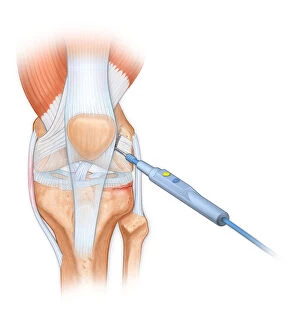
Background imageFibula Collection: Bovie used to cut through retincaculum, and clean up femur of Displaced patellar knee

Bovie used to cut through retincaculum, and clean up femur of Displaced patellar knee
Detail, Anatomy, Diagram, Muscle, Illustration, Bone, Cutting, Equipment, Injury, Medical, Tool, Biology, Femur, Surgery, Knee, Torn, Fractured, Meniscus, Cut Out, White Background, Vertical

Background imageFibula Collection: Illustration of the anterior knee, articular surface meniscus

Illustration of the anterior knee, articular surface meniscus
anatomy, body part, bone, cartilage, close-up, color image, femur, fibula, front view, human body part, human bone, human joint, human knee, illustration, joint - body part, knee, patella, meniscus

Background imageFibula Collection: Osteoarthritis of the knee, X-ray

Osteoarthritis of the knee, X-ray
Osteoarthritis of the knee, coloured X-ray

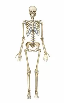
Background imageFibula Collection: Human skeletal system, front view

Human skeletal system, front view

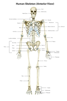
Background imageFibula Collection: Anterior view of human skeletal system, with labels

Anterior view of human skeletal system, with labels

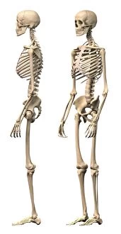
Background imageFibula Collection: Anatomy of male human skeleton, side view and perspective view

Anatomy of male human skeleton, side view and perspective view

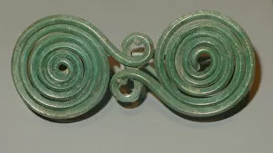
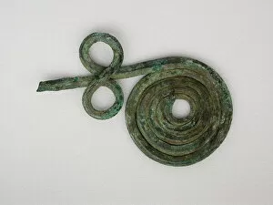
Background imageFibula Collection: Quatrefoil spiral fibula (garment pin), 7th century BCE. Creator: Unknown

Quatrefoil spiral fibula (garment pin), 7th century BCE. Creator: Unknown
Quatrefoil spiral fibula (garment pin), 7th century BCE

Background imageFibula Collection: Fibula Fragment, Geometric Period (800-600 BCE). Creator: Unknown

Fibula Fragment, Geometric Period (800-600 BCE). Creator: Unknown
Fibula Fragment, Geometric Period (800-600 BCE)

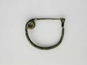
Background imageFibula Collection: Fibula (Garment Pin), Geometric Period (about 800 BCE). Creator: Unknown

Fibula (Garment Pin), Geometric Period (about 800 BCE). Creator: Unknown
Fibula (Garment Pin), Geometric Period (about 800 BCE)

Background imageFibula Collection: Bow Fibula, Geometric Period (800-600 BCE). Creator: Unknown

Bow Fibula, Geometric Period (800-600 BCE). Creator: Unknown
Bow Fibula, Geometric Period (800-600 BCE)

Background imageFibula Collection: Bow Fibula (wire), Geometric Period (800-600 BCE). Creator: Unknown

Bow Fibula (wire), Geometric Period (800-600 BCE). Creator: Unknown
Bow Fibula (wire), Geometric Period (800-600 BCE)

Background imageFibula Collection: Leech Fibula (Brooch), Geometric Period (800-700 BCE). Creator: Unknown

Leech Fibula (Brooch), Geometric Period (800-700 BCE). Creator: Unknown
Leech Fibula (Brooch), Geometric Period (800-700 BCE)

Background imageFibula Collection: Bow Fibula, Geometric Period (800-700 BCE). Creator: Unknown

Bow Fibula, Geometric Period (800-700 BCE). Creator: Unknown
Bow Fibula, Geometric Period (800-700 BCE)

Background imageFibula Collection: Spiral Fibula, Geometric Period (800-700 BCE). Creator: Unknown

Spiral Fibula, Geometric Period (800-700 BCE). Creator: Unknown
Spiral Fibula, Geometric Period (800-700 BCE)

Background imageFibula Collection: Fibula (leech type), Geometric Period (800-700 BCE). Creator: Unknown

Fibula (leech type), Geometric Period (800-700 BCE). Creator: Unknown
Fibula (leech type), Geometric Period (800-700 BCE)

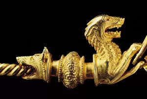
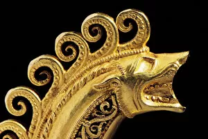
Background imageFibula Collection: Highly important La Tene warrior fibula, detail (gold) (see also 1065822-7)

Highly important La Tene warrior fibula, detail (gold) (see also 1065822-7)
1065824 Highly important La Tene warrior fibula, detail (gold) (see also 1065822-7) by Celtic, (3rd century BC); length: 14.1 cm; Private Collection; (add.info)

Background imageFibula Collection: Fibula in the form of a three-storey tripartite gate of rusticated masonry, c

Fibula in the form of a three-storey tripartite gate of rusticated masonry, c
483298 Fibula in the form of a three-storey tripartite gate of rusticated masonry, c.3rd - early 4th century AD (gilt silver) by Roman; length

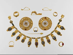
Background imageFibula Collection: Jewellery, early 5th century (gold and semi-precious stones)

Jewellery, early 5th century (gold and semi-precious stones)
3432856 Jewellery, early 5th century (gold and semi-precious stones) by Etruscan, (5th century BC); Metropolitan Museum of Art, New York

Background imageFibula Collection: Fibula, of a type common in Sweden and Norway (engraving)

Fibula, of a type common in Sweden and Norway (engraving)
3635886 Fibula, of a type common in Sweden and Norway (engraving) by English School, (19th century); Private Collection; (add.info.: Fibula, of a type common in Sweden and Norway)

Background imageFibula Collection: The Loan Art Collection in South Kensington Museum (engraving)

The Loan Art Collection in South Kensington Museum (engraving)
1588659 The Loan Art Collection in South Kensington Museum (engraving) by English School, (19th century); Private Collection; (add.info.: The Loan Art Collection in South Kensington Museum)

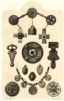
Background imageFibula Collection: Anglo-Saxon Relics, Personal Ornaments

Anglo-Saxon Relics, Personal Ornaments of Gold and Bronze. Enamelled ring of King Ethelwulf, enamelled brooch set with garnets, engraved pins, enamelled bronze cross with gold mount, gold cross

Background imageFibula Collection: Highly important La Tene warrior fibula, detail (gold) (see also 1065822-7)

Highly important La Tene warrior fibula, detail (gold) (see also 1065822-7)
1065823 Highly important La Tene warrior fibula, detail (gold) (see also 1065822-7) by Celtic, (3rd century BC); length: 14.1 cm; Private Collection; (add.info)

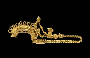
Background imageFibula Collection: Highly important La Tene warrior fibula (gold) (see also 1065823-7)

Highly important La Tene warrior fibula (gold) (see also 1065823-7)
1065822 Highly important La Tene warrior fibula (gold) (see also 1065823-7) by Celtic, (3rd century BC); length: 14.1 cm; Private Collection; (add.info)

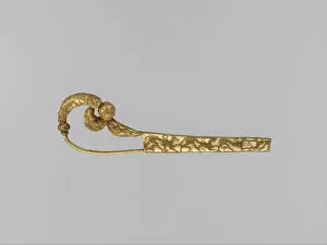
Background imageFibula Collection: Gold serpentine fibula (safety pin) with animals in granulation

Gold serpentine fibula (safety pin) with animals in granulation
3931606 Gold serpentine fibula (safety pin) with animals in granulation by Etruscan, (7th century BC); length: 8.9 cm; Metropolitan Museum of Art, New York, USA; Etruscan, out of copyright

Background imageFibula Collection: Radiate-Headed Brooch, from Marchelepot, Somme, probably Carolingian, c

Radiate-Headed Brooch, from Marchelepot, Somme, probably Carolingian, c
AMQ118633 Radiate-Headed Brooch, from Marchelepot, Somme, probably Carolingian, c.7th-10th century (gilt copper alloy) by Carolingian School; 8.6x4.6 cm; Ashmolean Museum, University of Oxford

Background imageFibula Collection: Jewellery from Camon, Amiens, Somme, Carolingian, 7th-10th century (silver

Jewellery from Camon, Amiens, Somme, Carolingian, 7th-10th century (silver
AMQ118627 Jewellery from Camon, Amiens, Somme, Carolingian, 7th-10th century (silver, gilded bronze) by Carolingian School; Ashmolean Museum, University of Oxford

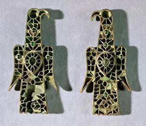
Background imageFibula Collection: Pair of Eagle-Shaped Brooches (metal and enamel)

Pair of Eagle-Shaped Brooches (metal and enamel)
PWI110914 Pair of Eagle-Shaped Brooches (metal and enamel) by Merovingian School; height: 14.2 cm; Musee National du Moyen Age et des Thermes de Cluny, Paris; ePeter Willi; French, out of copyright

Background imageFibula Collection: Fibula, from Palestrina with the inscription Manios (gold)

Fibula, from Palestrina with the inscription Manios (gold)
704094 Fibula, from Palestrina with the inscription Manios (gold) by Roman; Museo Preistorico e Etnographico Luigi Pigorini, Rome, Lazio, Italy; out of copyright

Background imageFibula Collection: Fibula, from the tomb of Bernardini di Palestrina (metal)

Fibula, from the tomb of Bernardini di Palestrina (metal)
704076 Fibula, from the tomb of Bernardini di Palestrina (metal) by Roman; Museo Nazionale Etrusco, Villa Giulia, Rome, Lazio, Italy; out of copyright

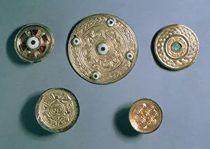
Background imageFibula Collection: Jewelled disc pendants, Anglo-Saxon (gold, silver gilt and semi-precious stones)

Jewelled disc pendants, Anglo-Saxon (gold, silver gilt and semi-precious stones)
AMQ118647 Jewelled disc pendants, Anglo-Saxon (gold, silver gilt and semi-precious stones) by Anglo-Saxon; Ashmolean Museum, University of Oxford, UK; eAshmolean Museum; English, out of copyright

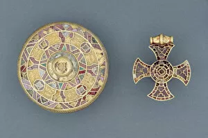
Background imageFibula Collection: Jewelled pendant cross, from Ixworth, Suffolk; and jewelled disc brooch, from Sarre, Kent

Jewelled pendant cross, from Ixworth, Suffolk; and jewelled disc brooch, from Sarre, Kent
AMQ118642 Jewelled pendant cross, from Ixworth, Suffolk; and jewelled disc brooch, from Sarre, Kent, Anglo-Saxon (gold, silver, garnets and shell) by Anglo-Saxon; length: 4.6 cm; Ashmolean Museum

Background imageFibula Collection: Jewelled disc brooch, from Sarre, Kent, Anglo-Saxon (gold with silver, garnets and shell)

Jewelled disc brooch, from Sarre, Kent, Anglo-Saxon (gold with silver, garnets and shell)
AMQ118639 Jewelled disc brooch, from Sarre, Kent, Anglo-Saxon (gold with silver, garnets and shell) by Anglo-Saxon; diam. 6.2 cm; Ashmolean Museum, University of Oxford


All Professionally Made to Order for Quick Shipping

The fibula, a slender bone in the human leg, plays a vital role in supporting the anatomy of the knee joint. Whether it's seen through an X-ray or depicted in artwork, this bone is often associated with various conditions and treatments. In cases where knee joint prosthesis is required, medical professionals rely on their expertise to restore mobility and alleviate pain. X-rays provide valuable insights into damaged knee ligaments, helping doctors determine the best course of action for recovery. Fractured ankles are another common occurrence that can be detected through X-rays. These images showcase the severity of such injuries and guide healthcare providers towards appropriate treatment options. To fully understand the complexity of our lower limbs, diagrams illustrating the bones of the right leg and hip offer a comprehensive view. The fibula stands alongside other crucial components like tibia and femur to ensure stability during movement. However, not all depictions revolve around ailments or injuries. Healthy knees captured through X-rays highlight how well-functioning joints should appear – a testament to proper care and maintenance. Artwork showcasing outer ankle ligaments sheds light on their significance in maintaining balance while inner ankle ligaments receive equal attention as they contribute to overall stability during physical activities. Sometimes extreme measures are necessary for patients suffering from severe knee issues; total knee replacement surgeries become essential interventions. Through x-rays post-surgery, one can observe how these procedures transform lives by restoring functionality. In unfortunate instances where broken legs require stabilization using pins or rods, medical intervention becomes paramount for successful healing processes. These interventions aim at aligning fractured bones accurately so that they can mend properly over time. Elephantiasis may seem unrelated but even this condition has its place when discussing fibulas - x-ray images reveal any potential complications caused by this disease affecting lymphatic vessels within extremities like legs. From anatomy lessons to surgical advancements and artistic representations – each aspect surrounding "fibula" provides unique perspectives on its role in our bodies.