Fraud Blocker Skip to main content

Flowering Part Collection

"Exploring the Intricate Beauty of Flowering Parts

Background imageFlowering Part Collection: Easter cactus stigma, SEM

Easter cactus stigma, SEM
Easter cactus stigma. Coloured scanning electron micrograph (SEM) of the stigma (pink) and style (green) of an Easter cactus flower (Rhipsalidopsis gaertneri)

Background imageFlowering Part Collection: Tea flower stamens, SEM

Tea flower stamens, SEM
Tea flower stamens. Coloured scanning electron micrograph (SEM) of the stamens of a flower of the tea plant (Camellia sp.). Stamens are the male reproductive structures of a flower

Background imageFlowering Part Collection: Buttercup flower, SEM

Buttercup flower, SEM
Buttercup flower (Ranunculus sp.), coloured scanning electron micrograph (SEM). The pistil (centre) is surrounded by the stamens (yellow)

Background imageFlowering Part Collection: Flower of the chickweed

Flower of the chickweed
False-colour scanning electron micrograph of the flower of common chickweed, Stellaria media, a self- pollinating plant found on uncultivated ground in Europe

Background imageFlowering Part Collection: Oil glands on petal of peppermint flower

Oil glands on petal of peppermint flower
Oil glands. Coloured scanning electron micrograph (SEM) of the surface of a corolla petal from the peppermint plant (Mentha piperita), showing oil (yellow) above its oil glands

Background imageFlowering Part Collection: Aubergine flower petal, SEM

Aubergine flower petal, SEM
Aubergine flower petal. Coloured scanning electron micrograph (SEM) of the surface of an aubergine (Solanum melongena) petal

Background imageFlowering Part Collection: Lily petal, SEM

Lily petal, SEM
Lily petal. Coloured scanning electron micrograph (SEM) of the surface of a lily (family Liliaceae). Magnification x58 at 6x7cm size

Background imageFlowering Part Collection: Columbine flower stamens, SEM

Columbine flower stamens, SEM
Columbine flower stamens. Coloured scanning electron micrograph (SEM) of the stamens (male sexual parts) of a columbine flower (Aquilegia sp.)

Background imageFlowering Part Collection: False-colour SEM of chickweed flower

False-colour SEM of chickweed flower
False-colour scanning electron micrograph of the reproductive structures of a flower of the chickweed, Stellaria media. The bulbous carpel, with the short-stalked female stigmas emerging from it

Background imageFlowering Part Collection: Yarrow flower petal, SEM

Yarrow flower petal, SEM
Yarrow flower petal. Coloured scanning electron micrograph (SEM) of a petal from the flower of a Japanese type of yarrow (Achillea alpina)

Background imageFlowering Part Collection: Periwinkle petal surface, SEM

Periwinkle petal surface, SEM
Periwinkle petal. Coloured scanning electron micrograph (SEM) of the surface of a periwinkle flower petal (family Apocynaceae)

Background imageFlowering Part Collection: Wild flower, SEM

Wild flower, SEM
Wild flower. Coloured scanning electron micrograph (SEM) of an unidentified flower. Magnification: x38 at 6x7cm size

Background imageFlowering Part Collection: Camellia flower stamens, SEM C015 / 9084

Camellia flower stamens, SEM C015 / 9084
Camellia flower stamens. Coloured scanning electron micrograph (SEM) of the stamens of a Camellia sp. flower. Stamens are the male reproductive structures of a flower

Background imageFlowering Part Collection: Orchid petal, SEM C016 / 0094

Orchid petal, SEM C016 / 0094
Orchid petal. Coloured scanning electron micrograph (SEM) of the surface of an orchid petal (Cymbidium sp.). It is covered in tiny epidermal hairs, or trichomes

Background imageFlowering Part Collection: Camellia flower stamens, SEM C015 / 9066

Camellia flower stamens, SEM C015 / 9066
Camellia flower stamens. Coloured scanning electron micrograph (SEM) of the stamens of a Camellia sp. flower. Stamens are the male reproductive structures of a flower

Background imageFlowering Part Collection: Orchid petal, SEM C016 / 0095

Orchid petal, SEM C016 / 0095
Orchid petal. Coloured scanning electron micrograph (SEM) of the surface of an orchid petal (Cymbidium sp.). It is covered in tiny epidermal hairs, or trichomes

Background imageFlowering Part Collection: Orchid petal, SEM C016 / 0097

Orchid petal, SEM C016 / 0097
Orchid petal. Coloured scanning electron micrograph (SEM) of the surface of an orchid petal (Cymbidium sp.). It is covered in tiny epidermal hairs, or trichomes

Background imageFlowering Part Collection: Orchid petal, SEM C016 / 0103

Orchid petal, SEM C016 / 0103
Orchid petal. Coloured scanning electron micrograph (SEM) of the surface of an orchid petal (Cymbidium sp.). It is covered in tiny epidermal hairs, or trichomes

Background imageFlowering Part Collection: Bellflower pistil, SEM C016 / 0064

Bellflower pistil, SEM C016 / 0064
Bellflower pistil. Coloured scanning electron micrograph (SEM) of the pistil of a bellflower plant (Campanula sp.). The pistil is the female reproductive part of a flower

Background imageFlowering Part Collection: Judas tree flower, SEM C016 / 0061

Judas tree flower, SEM C016 / 0061
Judas tree flower. Coloured scanning electron micrograph (SEM) of the reproductive parts of the flower of the Judas tree, or redbud (Cercis sp.)

Background imageFlowering Part Collection: Orchid petal, SEM C016 / 0098

Orchid petal, SEM C016 / 0098
Orchid petal. Coloured scanning electron micrograph (SEM) of the surface of an orchid petal (Cymbidium sp.). It is covered in tiny epidermal hairs, or trichomes

Background imageFlowering Part Collection: Bellflower pistil, SEM C016 / 0063

Bellflower pistil, SEM C016 / 0063
Bellflower pistil. Coloured scanning electron micrograph (SEM) of the pistil of a bellflower plant (Campanula sp.). The pistil is the female reproductive part of a flower

Background imageFlowering Part Collection: Judas tree flower, SEM C016 / 0062

Judas tree flower, SEM C016 / 0062
Judas tree flower. Coloured scanning electron micrograph (SEM) of the reproductive parts of the flower of the Judas tree, or redbud (Cercis sp.)

Background imageFlowering Part Collection: Orchid petal, SEM C016 / 0081

Orchid petal, SEM C016 / 0081
Orchid petal. Coloured scanning electron micrograph (SEM) of the surface of an orchid petal (Cymbidium sp.). It is covered in tiny epidermal hairs, or trichomes

Background imageFlowering Part Collection: Section through a lily ovary with ovules

Section through a lily ovary with ovules
Light micrograph of a transverse section through the ovary of a lily, Lilium sp.. Two ovules (female gametes) are visible within each of the three hollow chambers of the ovary

Background imageFlowering Part Collection: Hairs on sepal in rose flower

Hairs on sepal in rose flower
Rose sepal hairs. Coloured scanning electron micrograph (SEM) of the surface of a sepal in a Garden Rose flower Rosa sp. showing hairs

Background imageFlowering Part Collection: Lavender floret, SEM

Lavender floret, SEM
Lavender floret. Scanning electron micrograph (SEM) of a floret from a French lavender plant (Lavandula dentata). Individual florets are grouped together on stems to form the lavender flowers

Background imageFlowering Part Collection: Petal papillae in rose flower

Petal papillae in rose flower
Rose petal. Coloured scanning electron micrograph (SEM) showing papillae on the petal of a Garden Rose flower, Rosa sp. Papillae are projections from epidermal cells

Background imageFlowering Part Collection: SEM of pansy petal

SEM of pansy petal
Pansy petal. Coloured scanning electron micrograph (SEM) showing papillae on the petal of a pansy flower, Viola cornuta. Papillae are projections from epidermal cells

Background imageFlowering Part Collection: Stigmas of a Garden Rose flower

Stigmas of a Garden Rose flower
Rose stigmas. Coloured scanning electron micro- graph (SEM) of several stigmas in a Garden Rose flower, Rosa sp. The stigmas, with their numerous projections, form the free end of the carpel

Background imageFlowering Part Collection: Rose stamens showing pollen sacs

Rose stamens showing pollen sacs
Rose stamens. Coloured scanning electron micrograph (SEM) of stamens in a Garden Rose flower, Rosa sp. Stamens are the male reproductive organs of the flower

Background imageFlowering Part Collection: Petal papillae on a Snapdragon flower

Petal papillae on a Snapdragon flower
Petal papillae. Coloured scanning electron micrograph showing papillae on the petal of a Snapdragon flower, Antirrhinum majus. Papillae are projections from epidermal cells

Background imageFlowering Part Collection: Maize flower

Maize flower. Male flower of a maize plant (Zea mays), known as a tassel, with spikelets (green) and anthers (yellow). The anthers contain pollen

Background imageFlowering Part Collection: Flower anatomy, SEM

Flower anatomy, SEM
Flower anatomy. Coloured freeze-fracture scanning electron micrograph (SEM) of a flower with the top removed, showing a central ovary (pale pink, centre) containing ovules (green)

Background imageFlowering Part Collection: Lily bud, light micrograph

Lily bud, light micrograph
Lily bud. Light micrograph of a cross-section through an immature lily bud (Lilium sp.), showing the male and female reproductive organs of the flower

Background imageFlowering Part Collection: Lily anther, polar light micrograph

Lily anther, polar light micrograph
Lily anther. Polar light micrograph of a cross-section through a lily flower anther (Lilium sp.), showing four anther sacs (round structures)

Background imageFlowering Part Collection: Lily anther, light micrograph

Lily anther, light micrograph
Lily anther. Light micrograph of a cross-section through a lily flower anther (Lilium sp.), showing four anther sacs (curled structures)

Background imageFlowering Part Collection: Lily carpel, LM

Lily carpel, LM
Lily carpel. Light micrograph of a cross-section through a lily flower carpel (Lilium sp.), showing the three fused leaves that make up the pistil

Background imageFlowering Part Collection: Lily bud pistil and anthers, LM

Lily bud pistil and anthers, LM
Lily bud. Light micrograph of a cross-section through an immature lily bud (Lilium sp.), showing the male and female reproductive organs of the flower

Background imageFlowering Part Collection: Wallflower bud, light micrograph

Wallflower bud, light micrograph
Wallflower bud. Light micrograph of a cross-section through an immature wallflower bud (Cheiranthus cheiri), showing the male and female reproductive organs of the flower

Background imageFlowering Part Collection: Marsh marigold flower bud

Marsh marigold flower bud. Light micrograph of a cross-section through the anther of an immature marsh marigold (Caltha palustris) flower bud

Background imageFlowering Part Collection: Buttercup flower bud, light micrograph

Buttercup flower bud, light micrograph
Buttercup flower bud. Light micrograph of a cross-section through an immature wallflower bud (Ranunculus sp.), showing the male and female reproductive organs of the flower

Background imageFlowering Part Collection: Flower ovary and ovules, SEM

Flower ovary and ovules, SEM
Flower ovary and ovules. Coloured freeze-fracture scanning electron micrograph (SEM) through the ovary of a dicot flower. The ovary (purple) contains ovules (green)

Background imageFlowering Part Collection: Alexander flowers, light micrograph

Alexander flowers, light micrograph
Alexander flowers. Light micrograph of a section through a cluster of alexander flowers (Smyrnium olusatrum). Seen here are cross-sections of the central umbels

Background imageFlowering Part Collection: Cucumber flower bud, light micrograph

Cucumber flower bud, light micrograph
Cucumber flower bud. Light micrograph of a longitudinal section through the flower bud of a cucumber (Cucurbita sp.). The female pistil (red)

Background imageFlowering Part Collection: Snapdragon flower petal, SEM

Snapdragon flower petal, SEM
Snapdragon petal. Coloured scanning electron micrograph (SEM) of the surface of a snapdragon petal (Antirrhinum majus). The petal is covered in tiny epidermal hairs (trichomes)

Background imageFlowering Part Collection: Pansy flower, SEM

Pansy flower, SEM
Pansy flower reproductive parts. Coloured scanning electron micrograph (SEM) of reproductive parts from the flower of a pansy (Viola sp.) plant

Background imageFlowering Part Collection: Daisy fleabane flower, SEM

Daisy fleabane flower, SEM
Daisy fleabane flower. Coloured scanning electron micrograph (SEM) of the flower head of daisy fleabane (Erigeron sp.). This " flower" is actually a compound inflorescence composed of many


All Professionally Made to Order for Quick Shipping

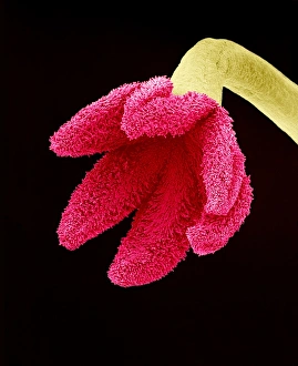
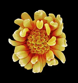
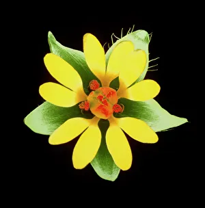
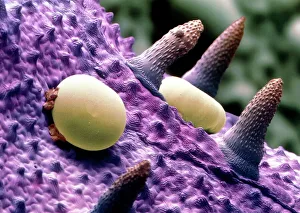
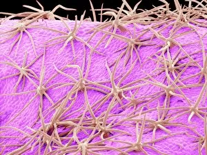
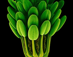
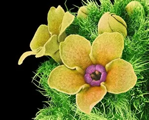
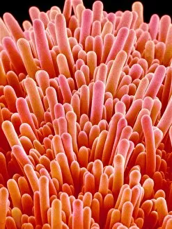
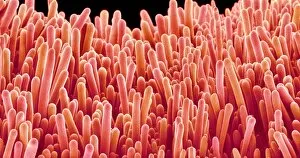
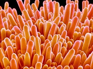
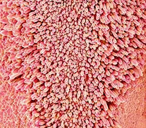
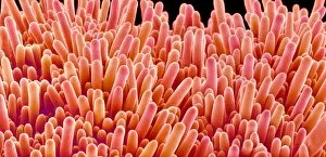
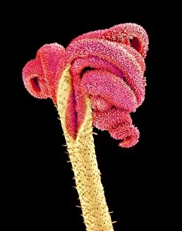
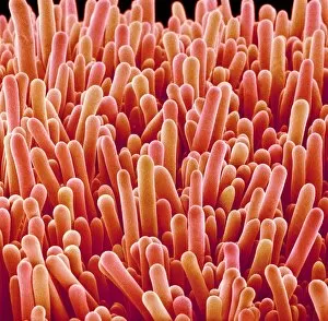
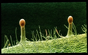
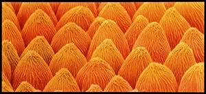
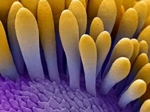
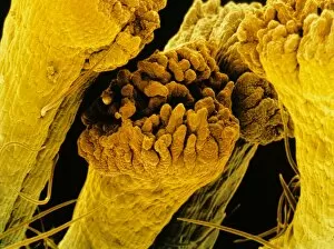
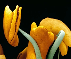
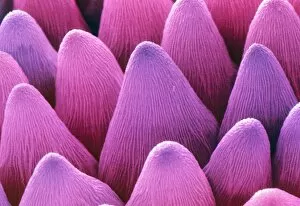
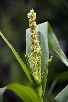
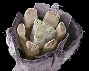
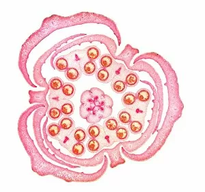
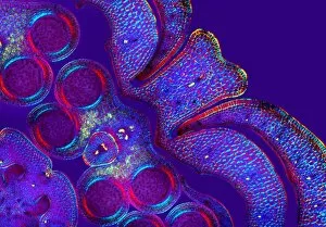
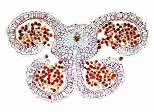
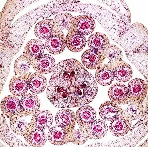
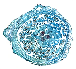
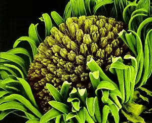
"Exploring the Intricate Beauty of Flowering Parts: A Captivating SEM Journey" Delve into the mesmerizing world of flowering parts as we embark on a captivating scanning electron microscope (SEM) adventure. Witness the intricate details and stunning structures that make up these botanical wonders. The Easter cactus stigma, magnified through SEM, reveals its delicate and feathery texture, showcasing nature's artistry at its finest. The Tea flower stamens come alive under the microscope, displaying their vibrant colors and fascinating patterns that attract pollinators with precision. A closer look at the Buttercup flower exposes its velvety petals in exquisite detail. Each petal seems to dance with light, inviting us to appreciate its ephemeral beauty. Meanwhile, the humble Chickweed flower surprises us with an unexpected burst of color when viewed through false-color SEM imaging techniques. Discovering hidden secrets within flowers continues as we observe oil glands on peppermint petals glistening like tiny droplets of gold. The Aubergine flower petal captivates our senses with its unique shape and intricate surface patterns revealed by SEM technology. Lily petals unfold before our eyes in breathtaking clarity under SEM examination; their graceful curves and delicate veins become works of art themselves. Columbine flower stamens reveal their complex arrangement, designed to ensure successful reproduction for this remarkable species. Diving deeper into nature's marvels, a false-color SEM image unveils the enchanting intricacies of a chickweed flower – each component playing a vital role in attracting pollinators while ensuring survival for generations to come. Yarrow's petal surface becomes an abstract masterpiece when observed through an electron microscope lens - every ridge and groove telling tales of resilience against harsh environments. And finally, witness the ovary of Opium poppy in astonishing detail; this microscopic view offers insights into how life begins within these magnificent blooms.