
Gas Exchange Collection
Gas exchange is a vital process that occurs in various organisms, enabling the exchange of gases between their internal systems and the environment


All Professionally Made to Order for Quick Shipping
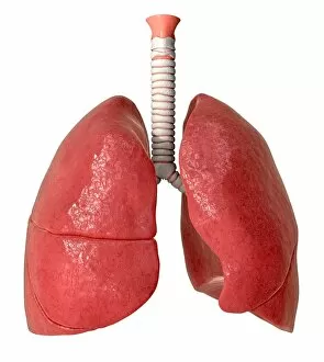
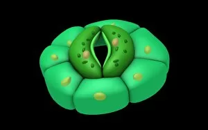
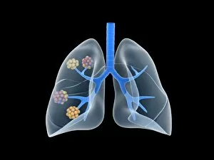
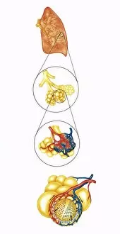
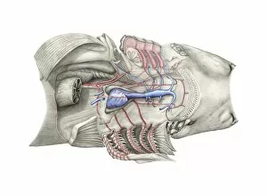
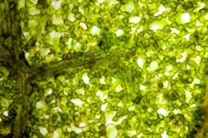
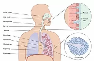
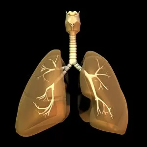
Gas exchange is a vital process that occurs in various organisms, enabling the exchange of gases between their internal systems and the environment. In plants, this intricate mechanism takes place through specialized structures like leaf pores. An English oak leaf pore, captured using scanning electron microscopy (SEM), showcases the intricacy of nature's design. Similarly, a French lavender leaf pore, also observed under SEM, reveals the remarkable complexity involved in gas exchange within plant tissues. These microscopic openings play a crucial role in facilitating the movement of gases such as oxygen and carbon dioxide. Moving from plants to animals, we delve into human anatomy to explore gas exchange on a different scale. The potato leaf stomata imaged using SEM provide an up-close view of these tiny openings found on leaves' surfaces. Stomata allow for gaseous interchange during photosynthesis while preventing excessive water loss. Shifting our focus to mammals' respiratory system, diseased alveoli in the lung serve as a stark reminder of how crucial proper gas exchange is for our health. When afflicted by diseases like pneumonia or emphysema, these delicate air sacs become inflamed or damaged impairing their ability to efficiently transfer oxygen into our bloodstream. Zooming even closer into our bodies at a cellular level brings us face-to-face with red blood cells captured via SEM. These biconcave-shaped cells are responsible for transporting oxygen throughout our body and removing waste products like carbon dioxide. To better understand how all these components work together harmoniously within human lungs, anatomical artwork provides an illustrative representation showcasing their interconnectedness and functionality. Conceptual images further aid us in visualizing both stomata and alveoli—the former depicting clusters of small pores on leaves' surfaces while the latter highlighting intricate air sacs within mammalian lungs. Through these conceptual representations, we can appreciate just how essential these structures are for efficient gas exchange across diverse organisms.