Fraud Blocker Skip to main content

Genetic Collection

"Unlocking the Secrets: Exploring the Fascinating World of Genetics" In this captivating journey, we delve into the intricate realm of genetics

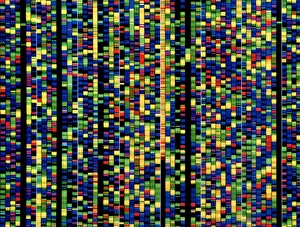
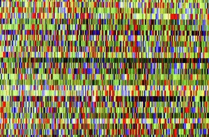
Background imageGenetic Collection: Computer screen showing a human genetic sequence

Computer screen showing a human genetic sequence
DNA sequencing. Computer screen showing a sequence of base pairs forming part of the human genetic code. The three billion base pairs that form the genetic code of human DNA (deoxyribonucleic acid)

Background imageGenetic Collection: DNA transcription, molecular model

DNA transcription, molecular model. Secondary structure of the enzyme RNA polymerase II synthesising a mRNA (messenger ribonucleic acid, lilac) strand from a DNA (deoxyribonucleic acid)

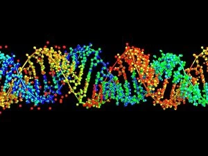
Background imageGenetic Collection: Double-stranded RNA molecule

Double-stranded RNA molecule. Computer model of the structure of double-stranded RNA (ribonucleic acid). The majority of RNA in a cell is in the single-stranded form

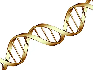
Background imageGenetic Collection: DNA molecule

DNA molecule, computer artwork. DNA (deoxyribonucleic acid) is composed of two strands twisted into a double helix. Each strand consists of a sugar-phosphate backbone attached to nucleotide bases

Background imageGenetic Collection: Leopard (Panthera pardus) black panther melanistic phase, adult, resting on log, captive

Leopard (Panthera pardus) black panther melanistic phase, adult, resting on log, captive

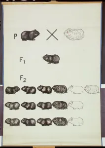
Background imageGenetic Collection: Poster demonstrating Mendels Law in Guinea Pigs (colour litho)

Poster demonstrating Mendels Law in Guinea Pigs (colour litho)
HMB306163 Poster demonstrating Mendels Law in Guinea Pigs (colour litho) by German School; 147x100 cm; Humboldt-Universitaet, Berlin

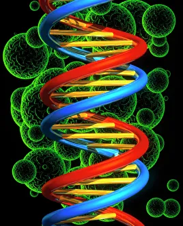
Background imageGenetic Collection: Computer artwork of a beta DNA segment and spheres

Computer artwork of a beta DNA segment and spheres
DNA molecule. Computer artwork of part of a strand of beta DNA (deoxyribonucleic acid) seen on a background of spheres. The spheres may represent bacteria

Background imageGenetic Collection: Nucleotide base matrix

Nucleotide base matrix. Computer artwork depicting a matrix of nucleotide bases: adenine (A), cytosine (C), guanine (G) and thymine (T)

Background imageGenetic Collection: DNA molecule, computer model

DNA molecule, computer model
DNA molecule. Computer artwork of the molecular structure of DNA (deoxyribonucleic acid). The DNA molecule is composed of two strands twisted into a double helix

Background imageGenetic Collection: Nucleosome molecule

Nucleosome molecule, computer model. A nucleosome is a subunit of chromatin, the substance that forms chromosomes. It consists of a short length of DNA (deoxyribonucleic acid)

Background imageGenetic Collection: DNA molecule, abstract image

DNA molecule, abstract image
DNA molecule. Abstract computer artwork of a view along the inside of a molecule of DNA (deoxyribonucleic acid). DNA contains sections called genes that encode the bodys genetic information

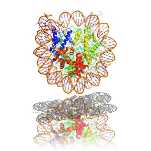
Background imageGenetic Collection: DNA nucleosome, molecular model

DNA nucleosome, molecular model
DNA nucleosome. Molecular model of a nucleosome, the fundamental repeating unit used to package DNA (deoxyribonucleic acid) inside cell nuclei

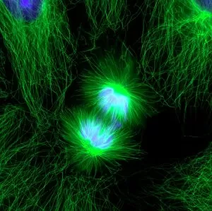
Background imageGenetic Collection: Cell division, fluorescent micrograph

Cell division, fluorescent micrograph
Cell division. Immunofluorescent light micrograph of a human epithelial cell (centre) during the late anaphase stage of mitosis

Background imageGenetic Collection: HIV reverse transcription enzyme

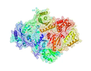
HIV reverse transcription enzyme. Molecular models of the reverse transcriptase enzyme found in HIV (the human immunodeficiency virus)

Background imageGenetic Collection: Gregor Mendel, Austrian botanist

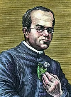
Gregor Mendel, Austrian botanist
Gregor Johann Mendel (1822-1884), Austrian botanist and founder of genetics. Mendel, the abbot of an abbey in Brno, carried out breeding experiments with pea plants (held in hand)

Background imageGenetic Collection: Mendels peas

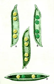
Mendels peas. Historical artwork of the peas (Pisum sp.) used by Gregor Mendel (1822-1884) in his experiments into heredity. He cross-bred peas that produced yellow (A) and green (B) peas

Background imageGenetic Collection: Fruit fly, SEM Z340 / 0699

Fruit fly, SEM Z340 / 0699
Fruit fly. Coloured scanning electron micrograph (SEM) of the head of a fruit fly (Drosophila busckii). Its two compound eyes (red) are seen on either side of the head

Background imageGenetic Collection: Zinc fingers bound to a DNA strand

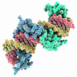
Zinc fingers bound to a DNA strand, molecular model. The double helix of DNA (deoxyribonucleic acid, red and yellow) is seen here with two Zif268 proteins (blue and green)

Background imageGenetic Collection: Full set of male chromosomes, SEM

Full set of male chromosomes, SEM
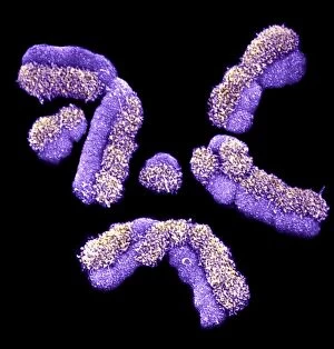
Set of human chromosomes, coloured scanning election micrograph (SEM). Chromosomes are a packaged form of the genetic material deoxyribonucleic acid (DNA)

Background imageGenetic Collection: Cystic fibrosis

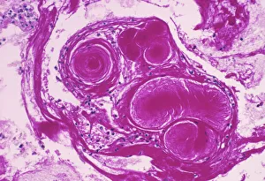
Cystic fibrosis. Light micrograph of a section through a bronchus (airway) of the lungs in a case of cystic fibrosis (mucoviscidosis)

Background imageGenetic Collection: Anti-cancer drug binding to DNA, AFM

Anti-cancer drug binding to DNA, AFM
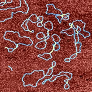
Drug-DNA complexes. Coloured atomic force micrograph (AFM) of plasmids (blue) of DNA (deoxyribonucleic acid) bound to the anti-cancer drug ditercalinium

Background imageGenetic Collection: DNA autoradiogram, artwork

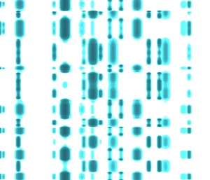
DNA autoradiogram, artwork. Autoradiograms show the order of nucleotide bases (basic building blocks) in a sample of DNA (deoxyribonucleic acid)

Background imageGenetic Collection: Computer artwork of DNA replication

Computer artwork of DNA replication
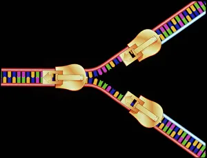
DNA replication. Computer artwork depicting DNA (deoxyribonucleic acid) replication. This segment of DNA is being " unzipped" to form a Y-shaped replication fork

Background imageGenetic Collection: Cell division, fluorescent micrograph

Cell division, fluorescent micrograph
Cell division. Immunofluorescent light micrograph of a human epithelial cell (centre) during the interphase stage of mitosis

Background imageGenetic Collection: Grapevine genome sequencing

Grapevine genome sequencing. Data from a gelelectrophoresis experiment to sequence the PinotNoir grape ( Vitis sp. ) genome

Background imageGenetic Collection: Fruit fly, SEM Z340 / 0700

Fruit fly, SEM Z340 / 0700
Fruit fly. Coloured scanning electron micrograph (SEM) of the head of a fruit fly (Drosophila busckii). Its two compound eyes (red) are seen on either side of the head

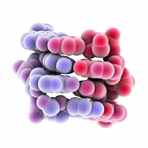
Background imageGenetic Collection: Z-DNA tetramer molecule C015 / 6557

Z-DNA tetramer molecule C015 / 6557
Z-DNA (deoxyribonucleic acid) tetramer, molecular model. DNA is composed of two strands twisted into a double helix. This is a tetramer of the molecule, containing four strands

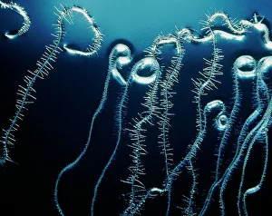
Background imageGenetic Collection: Roots of Thale cress plant, Arabidopsis thaliana

Roots of Thale cress plant, Arabidopsis thaliana
Root genetics research. Roots of the Thale cress plant, Arabidopsis thaliana, growing in a culture medium. The roots have fine root hairs growing horizontally out of them

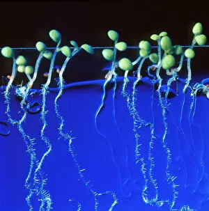
Background imageGenetic Collection: Cultured cress plant

Cultured cress plant
Plant genetics research. Thale cress plants, Arabidopsis thaliana, growing in a culture medium. The roots (lower frame) have fine root hairs growing horizontally out of them

Background imageGenetic Collection: Electrophoresis of RNA

Electrophoresis of RNA
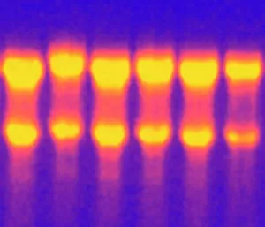
Liver RNA. Electrophoresis gel containing RNA (ribonucleic acid) isolated from liver tissue. The RNA molecules (white bands) are being observed under ultraviolet light

Background imageGenetic Collection: Shared DNA in humans and chimps, art

Shared DNA in humans and chimps, art
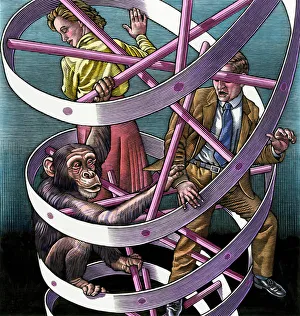
Shared DNA between humans and chimps, conceptual artwork. The humans look surprised to see the chimp so close to them in the double helix of DNA (deoxyribonucleic acid)

Background imageGenetic Collection: Mitochondrial DNA

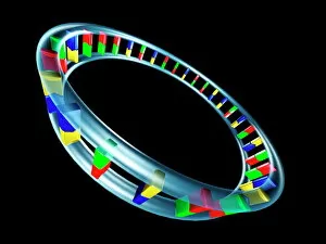
Mitochondrial DNA. Computer artwork of the genetic material (DNA, deoxyribonucleic acid) found in the cell structures called mitochondria

Background imageGenetic Collection: DNA analysis

DNA analysis, negative image. Graphs showing the results of DNA (deoxyribonucleic acid) sequencing. A DNA molecule consists of two sugar-phosphate backbones, arranged as a double helix

Background imageGenetic Collection: Human chromosomes, SEM C013 / 5002

Human chromosomes, SEM C013 / 5002
Human chromosomes. Coloured scanning electron micrograph (SEM) of human chromosomes prepared with the harlequin staining technique

Background imageGenetic Collection: Fragil Leaves

Fragil Leaves
Glass jar with green plants balancing on a stack of shards, creative spring concept. Dina Belenko

Background imageGenetic Collection: Picture No. 12479753

Picture No. 12479753
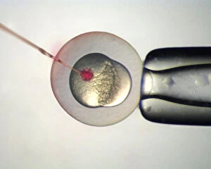
Microinjection of Zebrafish (Danio rerio) embryos Date:

Background imageGenetic Collection: DDE-90028007

DDE-90028007
North America, Canada, British Columbia, Princess Royal Island. Kermode (Spirit) Bear. Date: 22/10/2005

Background imageGenetic Collection: DDE-90028005

DDE-90028005
North America, Canada, British Columbia, Princess Royal Island. Kermode (Spirit) Bear. Date: 22/10/2005

Background imageGenetic Collection: DDE-90028006

DDE-90028006
North America, Canada, British Columbia, Princess Royal Island. Kermode (Spirit) Bear. Date: 22/10/2005

Background imageGenetic Collection: DDE-90028004

DDE-90028004
North America, Canada, British Columbia, Princess Royal Island. Kermode (Spirit) Bear. Date: 22/10/2005

Background imageGenetic Collection: DDE-90028003

DDE-90028003
North America, Canada, British Columbia, Princess Royal Island. Kermode (Spirit) Bear. Date: 22/10/2005

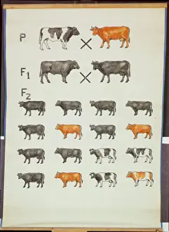
Background imageGenetic Collection: Poster demonstrating Mendels Law in Cattle (colour litho)

Poster demonstrating Mendels Law in Cattle (colour litho)
HMB306162 Poster demonstrating Mendels Law in Cattle (colour litho) by German School; 143x101 cm; Humboldt-Universitaet, Berlin

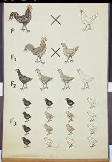
Background imageGenetic Collection: Poster demonstrating Mendels Law in Chickens (colour litho)

Poster demonstrating Mendels Law in Chickens (colour litho)
HMB306161 Poster demonstrating Mendels Law in Chickens (colour litho) by German School; Humboldt-Universitaet, Berlin, Germany; (add.info)

Background imageGenetic Collection: Portrait of Gregor Johann Mendel (b / w photo)

Portrait of Gregor Johann Mendel (b / w photo)
XEE4071019 Portrait of Gregor Johann Mendel (b/w photo) by German Photographer, (19th century); Private Collection; (add.info)

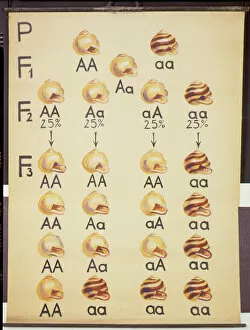
Background imageGenetic Collection: Poster of Cepea Snails (colour litho)

Poster of Cepea Snails (colour litho)
HMB306160 Poster of Cepea Snails (colour litho) by German School; 150x110 cm; Humboldt-Universitaet, Berlin, Germany; (add.info)

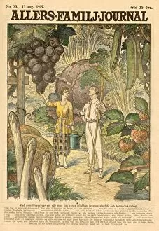
Background imageGenetic Collection: GIANT FRUIT

GIANT FRUIT
Thanks to genetic engineering, fruit and vegetables will be much bigger in the future... Date: 1919

Background imageGenetic Collection: Leucistic white lions (Panthera leo krugeri) male and juveniles, Captive

Leucistic white lions (Panthera leo krugeri) male and juveniles, Captive

Background imageGenetic Collection: Picture No. 12479765

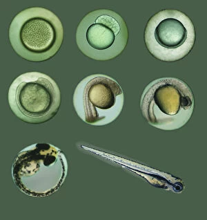
Picture No. 12479765
Zebrafish, Danio rerio, fry on aquarium. Since Date:


All Professionally Made to Order for Quick Shipping

"Unlocking the Secrets: Exploring the Fascinating World of Genetics" In this captivating journey, we delve into the intricate realm of genetics, where computer screens display mesmerizing human genetic sequences. The double-stranded RNA molecule stands as a testament to the complex nature of our genetic makeup. Witness DNA transcription in action through a stunning molecular model, unraveling the process that shapes our very existence. Amidst this exploration, an elegant leopard in its melanistic phase rests gracefully on a log, reminding us of the diversity and beauty found within genes. Computer artwork showcases a beta DNA segment surrounded by spheres, symbolizing both innovation and interconnectedness within our genetic code. The nucleotide base matrix unveils patterns that hold profound significance in understanding hereditary traits. As we peer into abstract images of DNA molecules, we are reminded of their remarkable structure and infinite possibilities they hold for life itself. The intricacies continue with the visualization of nucleosome molecules – tiny structures that play a crucial role in organizing our genetic material. Amidst these wonders lies an HIV reverse transcription enzyme; it serves as a stark reminder of how they can shape not only life but also disease. Yet even amidst challenges, there is hope as scientists tirelessly work to decipher these complexities and find solutions. Ultimately, this captivating journey through various facets of genetics leaves us awestruck by its elegance and complexity. It reminds us that every living being carries within them an extraordinary story written in their DNA – an ancient language connecting all forms of life on Earth.