
Genetics Collection
"Unlocking the Secrets of Life: Exploring the Fascinating World of Genetics" From the intricate DNA molecule to the X and Y chromosomes


All Professionally Made to Order for Quick Shipping
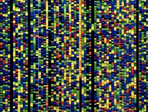
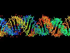
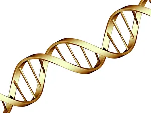
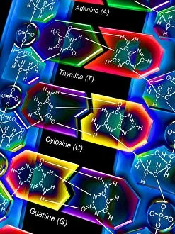
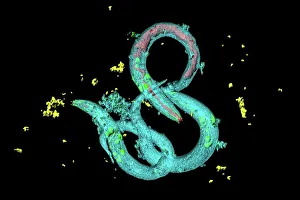
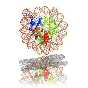
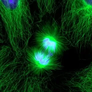
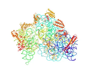
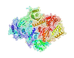
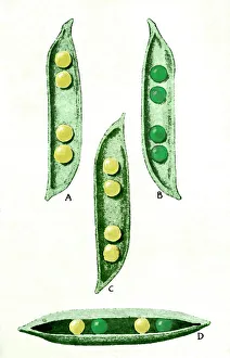
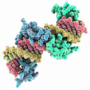
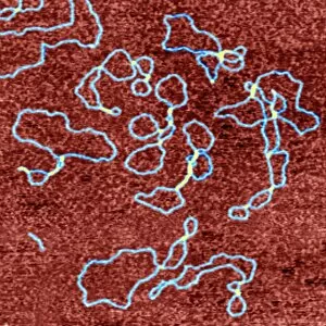
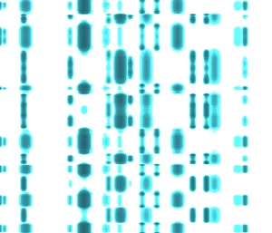
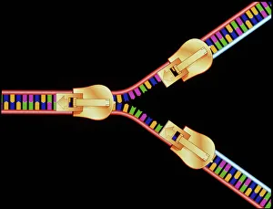
"Unlocking the Secrets of Life: Exploring the Fascinating World of Genetics" From the intricate DNA molecule to the X and Y chromosomes, a captivating field that unravels the blueprint of life. As we peer into a computer screen displaying a human genetic sequence, we witness the complexity encoded within our very cells. The double-stranded RNA molecule serves as a messenger, carrying vital information for DNA transcription. Molecular models illustrate how this process shapes our traits and characteristics. It was through their groundbreaking work that Watson and Crick discovered the structure of DNA, forever changing our understanding of genetics. Richard Dawkins, an esteemed British science writer, has played an influential role in popularizing genetics among masses. His insightful writings have shed light on evolutionary biology and its connection to our genetic makeup. Intriguingly captured by scanning electron microscopy (SEM), an embryonic stem cell alongside a needle reminds us of the immense potential held within these tiny building blocks. Mitosis comes alive under a light micrograph, showcasing how cells divide and multiply with precision. Computer artwork depicting beta DNA segments interlaced with spheres hints at ongoing research pushing boundaries in genetic engineering. The nucleotide base matrix acts as a foundation for decoding genetic information - each letter representing crucial instructions embedded within our genes. Genetics holds endless possibilities - from unraveling hereditary diseases to designing personalized medicine based on individual genomes. With every discovery made in this ever-evolving field, humanity inches closer towards harnessing nature's codebook for better health and understanding ourselves more deeply than ever before.