Fraud Blocker Skip to main content

Glands Collection

"Glands: Nature's Hidden Gems Unveiled" Botanik Digitalis purpurea L. Fingerhut 160: 1 - Exploring the intricate world of plant glands

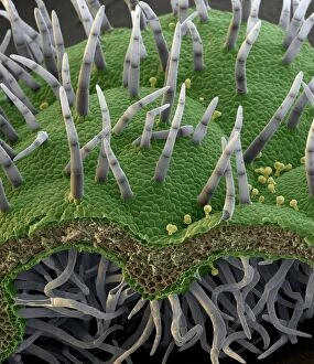
Background imageGlands Collection: Botanik Digitalis purpurea L. Fingerhut 160: 1

Botanik Digitalis purpurea L. Fingerhut 160: 1
Foxglove leaf. Coloured scanning electron micrograph (SEM) of a section through a leaf from a foxglove (Digitalis purpurea) plant, showing its hairs (trichomes)

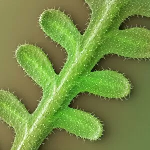
Background imageGlands Collection: French lavender leaf surface, SEM

French lavender leaf surface, SEM
French lavender leaf surface. Coloured scanning electron micrograph (SEM) of a section of French lavender (Lavandula dentata) leaf, showing the tooth-like structures (trichomes)

Background imageGlands Collection: Leaf oil glands, SEM

Leaf oil glands, SEM
Leaf oil glands. Coloured scanning electron micrograph (SEM) of a thyme plants leaf surface (Thymus vulgaris). Two types of leaf trichomes (hairs) are seen

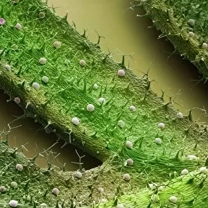
Background imageGlands Collection: French lavender leaf, SEM

French lavender leaf, SEM
French lavender (Lavandula dentata) leaf. Coloured scanning electron micrograph (SEM) of a section of a French lavender leaf, showing the tooth-like structures (trichomes)

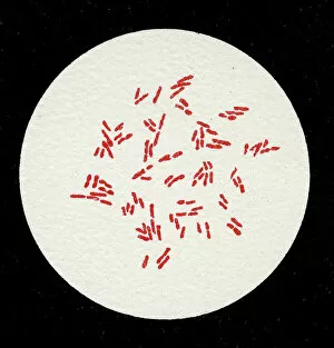
Background imageGlands Collection: Colony of Bacterium mallei, 1906 (litho)

Colony of Bacterium mallei, 1906 (litho)
3501906 Colony of Bacterium mallei, 1906 (litho) by German School, (20th century); Private Collection; (add.info.: Bacteria constitute a large domain of prokaryotic microorganisms)

Background imageGlands Collection: DDE-90036392

DDE-90036392
Luxembourg, Luxembourg City. Les Trois Glands (The Three Acorns) fortress with the Modern Art Museum in the background. Date: 14/10/2005

Background imageGlands Collection: Frontal View of the Muscles and Glands of the Human Neck, 1906 (engraving)

Frontal View of the Muscles and Glands of the Human Neck, 1906 (engraving)
2622229 Frontal View of the Muscles and Glands of the Human Neck, 1906 (engraving) by German School, (20th century); Private Collection; (add.info)

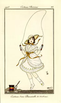
Background imageGlands Collection: Girl skipping rope at the beach in white dress

Girl skipping rope at the beach in white dress and bonnet decorated with flowers. Costume d une damoiselle de 6 a 8 ans. Handcoloured pochoir (stencil)

Background imageGlands Collection: Woman in the Crown or Couronne hairdo, 1783

Woman in the Crown or Couronne hairdo, 1783
Woman in the Crown or Couronne hairdo, a backcombed hairstyle surmounted by a pouf hat with pleats, pearls and tassels. Tape renverse surmonte d un Pouf a plis ronds avec des perles et 2 Glands dit a

Background imageGlands Collection: Still Life with Acorns and Horse Chestnuts (w / c, b / c & graphite on paper)

Still Life with Acorns and Horse Chestnuts (w / c, b / c & graphite on paper)
XYC284411 Still Life with Acorns and Horse Chestnuts (w/c, b/c & graphite on paper) by English School, (19th century); 21.7x28.6 cm; Yale Center for British Art, Paul Mellon Collection, USA; English

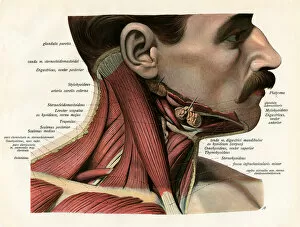
Background imageGlands Collection: Lateral View of the Muscles and Glands of the Human Neck, 1906 (engraving)

Lateral View of the Muscles and Glands of the Human Neck, 1906 (engraving)
2622230 Lateral View of the Muscles and Glands of the Human Neck, 1906 (engraving) by German School, (20th century); Private Collection; (add.info)

Background imageGlands Collection: The salivary glands (engraving)

The salivary glands (engraving)
3634175 The salivary glands (engraving) by English School, (19th century); Private Collection; (add.info.: The salivary glands)

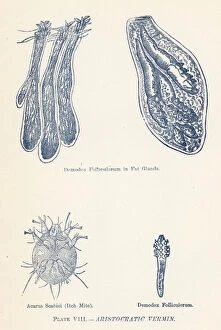
Background imageGlands Collection: Aristocratic Vermin (colour litho)

Aristocratic Vermin (colour litho)
2799335 Aristocratic Vermin (colour litho) by English School, (19th century); Private Collection; (add.info.: Aristocratic Vermin)

Background imageGlands Collection: Perspiration, sweat glands from skin of thumb (litho)

Perspiration, sweat glands from skin of thumb (litho)
6013660 Perspiration, sweat glands from skin of thumb (litho) by English School, (20th century); Private Collection; (add.info.: Perspiration, sweat glands from skin of thumb)

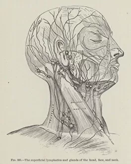
Background imageGlands Collection: The superficial lymphatics and glands of the head, face, and neck (engraving)

The superficial lymphatics and glands of the head, face, and neck (engraving)
3634187 The superficial lymphatics and glands of the head, face, and neck (engraving) by English School, (19th century); Private Collection; (add.info)

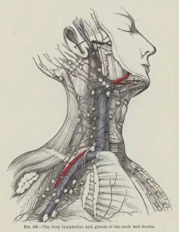
Background imageGlands Collection: The deep lymphatics and glands of the neck and thorax (engraving)

The deep lymphatics and glands of the neck and thorax (engraving)
3634185 The deep lymphatics and glands of the neck and thorax (engraving) by English School, (19th century); Private Collection; (add.info.: The deep lymphatics and glands of the neck and thorax)

Background imageGlands Collection: The Skin (colour litho)

The Skin (colour litho)
2799331 The Skin (colour litho) by English School, (19th century); Private Collection; (add.info.: The Skin. Illustration for The Home Handbook of Domestic Hygiene)

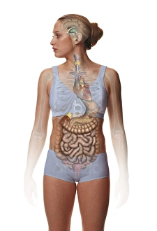
Background imageGlands Collection: Woman standing, body facing forward head turned to one side, illustration overlay showing skeleton

Woman standing, body facing forward head turned to one side, illustration overlay showing skeleton and inner organs

Background imageGlands Collection: NA

NA
Closeup of a thunbergia sky flower, Thunbergia battiscombei

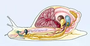
Background imageGlands Collection: Cross-section of a Giant African land snail (Achatina)

Cross-section of a Giant African land snail (Achatina)

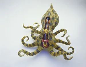
Background imageGlands Collection: Model of Blue-ringed Octopus with legs curling, cross-section showing orange venom glands and duct

Model of Blue-ringed Octopus with legs curling, cross-section showing orange venom glands and duct, blue crop storing undigested food, large yellow brain, red mouth, salivary glands and tongue

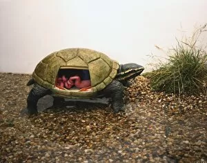
Background imageGlands Collection: Side view of model of Stinkpot Turtle crawling from water with head and legs extending

Side view of model of Stinkpot Turtle crawling from water with head and legs extending, scutes covering bony shell, section of shell removed showing internal organs including red stink glands

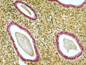
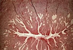
Background imageGlands Collection: Cervix, light micrograph F006 / 9805

Cervix, light micrograph F006 / 9805
Cervix. Light micrograph of a section though the mucosa of the cervix, the neck of the uterus. The purple-lined areas are cervical glands, which produce mucus (pink inside the glands)
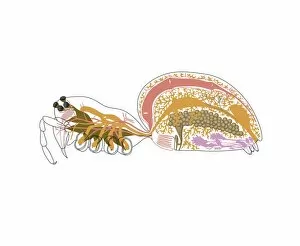
Background imageGlands Collection: Spider anatomy, artwork

Spider anatomy, artwork
Spider anatomy. Computer artwork showing the main organs of a typical female spider
Background imageGlands Collection: Fight-or-flight response, artwork

Fight-or-flight response, artwork
Fight-or-flight response. Artwork showing the bodys fight-or-flight response to a threat. Visual perception (yellow arrow) can trigger fear memories controlled by the brains amygdala (yellow)

Background imageGlands Collection: Trachea lining, SEM C013 / 7126

Trachea lining, SEM C013 / 7126
Trachea lining. Coloured scanning electron micrograph (SEM) of the lining of a trachea (wind pipe), showing the cilia (hair-like) and goblet cells (orange). The trachea links the larynx to the lungs

Background imageGlands Collection: Trachea lining, SEM C013 / 7122

Trachea lining, SEM C013 / 7122
Trachea lining. Coloured scanning electron micrograph (SEM) of a section through the wall of a trachea (wind pipe). The trachea links the larynx to the lungs

Background imageGlands Collection: Skin and hair follicles, light micrograph

Skin and hair follicles, light micrograph
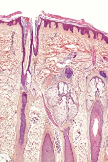
Skin and hair follicles. Light micrograph of a section through skin, showing hair follicles (dark purple circular structures). Haematoxylin and eosin stain

Background imageGlands Collection: Parathyroid hormone molecule

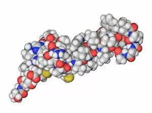
Parathyroid hormone molecule. Computer model showing the structure of parathyroid hormone (PTH), or parathormone. Atoms are colour-coded (carbon: dark grey, hydrogen: light grey, oxygen: red)

Background imageGlands Collection: Moon jellyfish sex glands

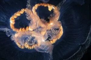
Moon jellyfish sex glands. Close up of the sex glands of an ephyra medusa of the cnidarian Aurelia aurita. Medusae are the sexually mature

Background imageGlands Collection: Luxembourg - Fort Thungen - Drai Eechelen Park

Luxembourg - Fort Thungen - Drai Eechelen Park
Luxembourg - Fort Thungen - a historic fortification sited in Drai Eechelen Park, in the Kirchberg quarter. It is also colloquially known as Three Acorns ( Trois Glands )

Background imageGlands Collection: Glands of Stomach in Birds

Glands of Stomach in Birds

Background imageGlands Collection: Picture No. 10776588

Picture No. 10776588
Dog - being examined by vet - emptying anal glands Date:

Background imageGlands Collection: Picture No. 10896292

Picture No. 10896292
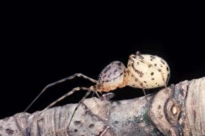
Spider - Its huge cephalothorax contains salivary glands it uses to lime preys (Scytodes thoracica) Date:

Background imageGlands Collection: Overhead view of model of upper torso of Giant Tiger Centipede

Overhead view of model of upper torso of Giant Tiger Centipede, pair of legs attached to each segment, venom claws behind antennae, cross-section showing venom glands with duct, nerve and muscle

Background imageGlands Collection: Front-on view of the head of a Red Spitting Cobra showing the venom glands situated below the eyes

Front-on view of the head of a Red Spitting Cobra showing the venom glands situated below the eyes. The broadest part of the head is well behind the eyes, narrowing sharply towards the snout

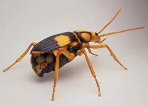
Background imageGlands Collection: Side view of model of black and yellow Bombardier Beetle with yellow legs

Side view of model of black and yellow Bombardier Beetle with yellow legs, cross section showing venom glands and reservoir, explosion chamber filled with red liquid with one-way valve, side view

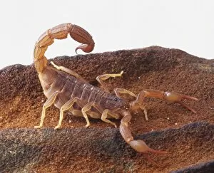
Background imageGlands Collection: Desert Scorpion, strong pincers for catching food, sandy colouring for camouflage

Desert Scorpion, strong pincers for catching food, sandy colouring for camouflage, segmented tail arching over, sting with swollen poison glands

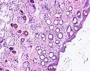
Background imageGlands Collection: Human skin section, light micrograph

Human skin section, light micrograph
Human skin. Light micrograph of a section through healthy human skin. The outer surface of the skin is at top. The uppermost surface is the epidermis

Background imageGlands Collection: Human skin section, light micrograph P710 / 0472

Human skin section, light micrograph P710 / 0472
Human skin. Light micrograph of a section through healthy human skin. The outer surface of the skin is at top. The uppermost surface is the epidermis

Background imageGlands Collection: Houttuynia cordata leaf surface, SEM C015 / 5078

Houttuynia cordata leaf surface, SEM C015 / 5078
Leaf surface. Coloured environmental scanning electron micrograph (ESEM) of two trichomes (hair-like) on a Houttuynia cordata leaf. These are glandular trichomes, which produce an orange-scented oil

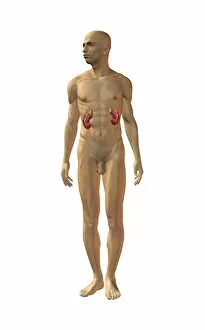
Background imageGlands Collection: Kidneys, anatomical artwork

Kidneys, anatomical artwork
Kidneys. Anatomical computer artwork of a mans body, showing the position of the kidneys (red, centre left and right) and adrenal glands (beige)

Background imageGlands Collection: Ischaemic bowel, light micrograph

Ischaemic bowel, light micrograph
Ischaemic bowel. Light micrograph of a section through a portion of intestine that has been damaged by an interruption of the blood supply, or ischaemia

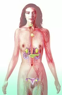
Background imageGlands Collection: Female endocrine glands, artwork C016 / 7522

Female endocrine glands, artwork C016 / 7522
Female endocrine glands. Computer artwork showing the location of the endocrine glands (yellow) in a womans body. These are (from top to bottom): thyroid, thymus, adrenal glands (x2), pancreas

Background imageGlands Collection: Insect defence glands, artwork C016 / 7681

Insect defence glands, artwork C016 / 7681
Insect defence glands. Computer artwork of a beetle (order Coleoptera, left) and ant (family Formicidae, right), showing the location of the glands that secrete defensive chemicals

Background imageGlands Collection: Cervix, light micrograph

Cervix, light micrograph
Cervix. Light micrograph of a section through the mucosa of the cervix, the neck of the uterus. This is the endocervix, which borders the uterus. It is lined with columnar epithelial cells

Background imageGlands Collection: Enteroendocrine cell, TEM

Enteroendocrine cell, TEM
Enteroendocrine cell. Transmission electron micrograph (TEM) of a section through an enteroendocrine cell within a gland of the stomach

Background imageGlands Collection: Colon polyp, light micrograph C016 / 0512

Colon polyp, light micrograph C016 / 0512
Colon polyp. Light micrograph of a section through part of a tubulovillous adenoma of the mucosa of the large bowel (colon)


All Professionally Made to Order for Quick Shipping

"Glands: Nature's Hidden Gems Unveiled" Botanik Digitalis purpurea L. Fingerhut 160: 1 - Exploring the intricate world of plant glands. French lavender leaf surface, SEM - A closer look at the fascinating structures on a lavender leaf. Leaf oil glands, SEM - Unlocking the secrets behind essential oils through microscopic examination. Orange fruit, light micrograph - Delving into the hidden wonders of an orange's glandular system. Dog anatomy, artwork - Discovering how glands play a vital role in our furry friends' physiology. French lavender leaf surface, SEM (repeated) - Peering into the mesmerizing patterns on a lavender leaf once more. Colony of Bacterium mallei, 1906 (litho) - Examining bacterial colonies and their impact on glandular health. DDE-90036392 – Unraveling the mysteries surrounding specific gland-related research or findings. Frontal View of the Muscles and Glands of Human Neck, 1906 (engraving) – Appreciating our complex neck anatomy and its glandular intricacies throughout history. Girl skipping rope at the beach in white dress – Amidst all scientific exploration lies simple joys like skipping ropes; reminding us that even within ourselves lie remarkable glands waiting to be discovered. From plants to animals and humans alike, these captivating glimpses into various aspects of "glands" shed light on their significance in nature's grand design while reminding us that beauty can be found even in life's tiniest details.